The DRP1 Antibody (CAB2586) is a high-quality antibody developed for reliable detection and analysis of target proteins. This antibody, raised in rabbits, is highly specific to human samples and is validated for use in Western blot applications. By binding to the DNM1L protein, this antibody enables accurate detection and analysis in various cell types, making it ideal for investigations in the fields of cell biology, neurology, and metabolism.DNM1L, also known as dynamin-1-like protein, plays a crucial role in maintaining mitochondrial health and function by regulating organelle division and clearance.
This antibody is validated for use in WB, IHC-P, IF/ICC, ELISA applications and has demonstrated reactivity against Human, Mouse, Rat samples.
Product Name:
DRP1 Antibody
SKU:
CAB2586
Size:
20μL, 100μL
Reactivity:
Human, Mouse, Rat
Conjugate:
Unconjugated
Immunogen:
Recombinant protein (or fragment).This information is considered to be commercially sensitive.
This gene encodes a member of the dynamin superfamily of GTPases. The encoded protein mediates mitochondrial and peroxisomal division, and is involved in developmentally regulated apoptosis and programmed necrosis. Dysfunction of this gene is implicated in several neurological disorders, including Alzheimer's disease. Mutations in this gene are associated with the autosomal dominant disorder, encephalopathy, lethal, due to defective mitochondrial and peroxisomal fission (EMPF). Alternative splicing results in multiple transcript variants encoding different isoforms.
Purification Method
Affinity purification
Gene ID
10059
RRID
AB_2764472
Buffer Information
Store at -20℃. Avoid freeze / thaw cycles. Buffer: PBS with 0.01% thimerosal,50% glycerol,pH7.3.
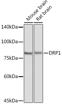
Western blot analysis of various lysates using DRP1 Rabbit pAb (CAB2586) at 1:8000 dilution. Secondary antibody: HRP-conjugated Goat anti-Rabbit IgG (H+L) (CABS014) at 1:10000 dilution. Lysates/proteins: 25μg per lane. Blocking buffer: 3% nonfat dry milk in TBST. Detection: ECL Basic Kit (AbGn00020). Exposure time: 1s.
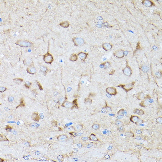
Immunohistochemistry analysis of paraffin-embedded Rat brain using DRP1 Rabbit pAb (CAB2586) at dilution of 1:200 (40x lens). High pressure antigen retrieval performed with 0.01M Citrate buffer (pH 6.0) prior to IHC staining.
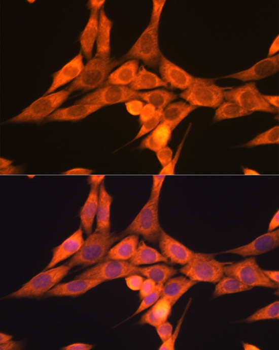
Immunofluorescence analysis of NIH/3T3 cells using DRP1 Rabbit pAb (CAB2586) at dilution of 1:100 (40x lens). Secondary antibody: Cy3-conjugated Goat anti-Rabbit IgG (H+L) (CABS007) at 1:500 dilution. Blue: DAPI for nuclear staining.