The Dopamine Receptor D3 (DRD3) Antibody (CAB14622) is a high-quality antibody developed for reliable detection and analysis of target proteins. This antibody, generated in rabbits, exhibits high reactivity with human samples and has been validated for use in Western blot applications. By binding to the DRD3 protein, this antibody enables the detection and analysis of DRD3 in various cell types, making it an ideal tool for studying the role of DRD3 in neurological disorders and psychiatric conditions.
This antibody is validated for use in WB, ELISA applications and has demonstrated reactivity against Mouse, Rat samples.
Product Name:
Dopamine Receptor D3 (DRD3) Antibody
SKU:
CAB14622
Size:
20μL, 100μL
Reactivity:
Mouse, Rat
Conjugate:
Unconjugated
Immunogen:
Synthetic peptide. This information is considered to be commercially sensitive.
Recommended starting concentration is 1 μg/mL. Please optimize the concentration based on your specific assay requirements.
Synonyms:
D3DR, ETM1, FET1, Dopamine Receptor D3 (DRD3)
Positive Sample:
Mouse brain, Rat brain
Cellular Localization:
Cell Membrane, Multi-Pass Membrane Protein.
Calculated MW:
44kDa
Observed MW:
44kDa
This gene encodes the D3 subtype of the five (D1-D5) dopamine receptors. The activity of the D3 subtype receptor is mediated by G proteins which inhibit adenylyl cyclase. This receptor is localized to the limbic areas of the brain, which are associated with cognitive, emotional, and endocrine functions. Genetic variation in this gene may be associated with susceptibility to hereditary essential tremor 1. Alternative splicing of this gene results in transcript variants encoding different isoforms, although some variants may be subject to nonsense-mediated decay (NMD).
Purification Method
Affinity purification
Gene ID
1814
RRID
AB_2761496
Buffer Information
Store at -20℃. Avoid freeze / thaw cycles. Buffer: PBS with 0.01% thimerosal,50% glycerol,pH7.3.
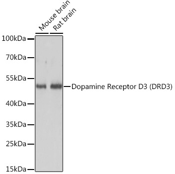
Western blot analysis of various lysates using Dopamine Receptor D3 (DRD3) Rabbit pAb (CAB14622) at 1:3000 dilution. Secondary antibody: HRP-conjugated Goat anti-Rabbit IgG (H+L) (CABS014) at 1:10000 dilution. Lysates/proteins: 25μg per lane. Blocking buffer: 3% nonfat dry milk in TBST. Detection: ECL Basic Kit (AbGn00020). Exposure time: 1s.


 CLIA Kit (RTES00185)")

 ELISA Kit (AEKE00318)")