The DROSHA Monoclonal Antibody (CAB19598) is a high-quality antibody developed for reliable detection and analysis of target proteins. This antibody, raised in rabbits, is suitable for use in various applications including Western blot, immunofluorescence, and immunohistochemistry.Drosha plays a crucial role in the biogenesis of microRNAs, which are small non-coding RNAs that regulate gene expression. Dysregulation of microRNA processing has been implicated in various diseases, including cancer, neurodegenerative disorders, and cardiovascular diseases.
This antibody is validated for use in WB, IF/ICC, ELISA applications and has demonstrated reactivity against Human samples.
Product Name:
DROSHA Monoclonal Antibody
SKU:
CAB19598
Size:
20μL, 100μL
Reactivity:
Human
Clone Number:
ARC0077
Conjugate:
Unconjugated
Immunogen:
Synthetic peptide. This information is considered to be commercially sensitive.
This gene encodes a ribonuclease (RNase) III double-stranded RNA-specific ribonuclease and subunit of the microprocessor protein complex, which catalyzes the initial processing step of microRNA (miRNA) synthesis. The encoded protein cleaves the stem loop structure from the primary microRNA (pri-miRNA) in the nucleus, yielding the precursor miRNA (pre-miRNA), which is then exported to the cytoplasm for further processing. In a human cell line lacking a functional copy of this gene, canonical miRNA synthesis is reduced. Somatic mutations in this gene have been observed in human patients with kidney cancer.
Purification Method
Affinity purification
Gene ID
29102
RRID
AB_2862691
Buffer Information
Store at -20℃. Avoid freeze / thaw cycles. Buffer: PBS containing 50% glycerol and 0.05% BSA, preserved with proclin300 or sodium azide, pH 7.3.
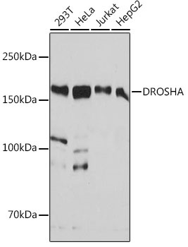
Western blot analysis of various lysates using DROSHA Rabbit mAb (CAB19598) at 1:1000 dilution. Secondary antibody: HRP-conjugated Goat anti-Rabbit IgG (H+L) (CABS014) at 1:10000 dilution. Lysates/proteins: 25μg per lane. Blocking buffer: 3% nonfat dry milk in TBST. Detection: ECL Basic Kit (AbGn00020). Exposure time: 60s.
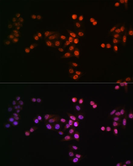
Immunofluorescence analysis of HeLa cells using DROSHA Rabbit mAb (CAB19598) at a dilution of 1:100 (40x lens). Secondary antibody: Cy3-conjugated Goat anti-Rabbit IgG (H+L)(CABS007) at 1:500 dilution. Blue: DAPI for nuclear staining.