The ECH1 Antibody (CAB12944) is a high-quality antibody developed for reliable detection and analysis of target proteins. This antibody, raised in rabbits, exhibits high reactivity with human samples and is validated for use in Western blot applications. By binding to the ECH1 protein, this antibody enables accurate detection and analysis in various cell types, making it an ideal tool for studies in metabolism, lipid metabolism disorders, and related diseases.ECH1 plays a crucial role in fatty acid oxidation, a process essential for energy production and maintaining metabolic balance.
This antibody is validated for use in WB, IHC-P, ELISA applications and has demonstrated reactivity against Human, Mouse, Rat samples.
Product Name:
ECH1 Antibody
SKU:
CAB12944
Size:
20μL, 100μL
Reactivity:
Human, Mouse, Rat
Conjugate:
Unconjugated
Immunogen:
Recombinant protein (or fragment).This information is considered to be commercially sensitive.
Recommended starting concentration is 1 μg/mL. Please optimize the concentration based on your specific assay requirements.
Synonyms:
HPXEL, ECH1
Positive Sample:
Hep G2, Rat heart, Mouse heart
Cellular Localization:
Mitochondrion, Peroxisome.
Calculated MW:
36kDa
Observed MW:
36kDa
This gene encodes a member of the hydratase/isomerase superfamily. The gene product shows high sequence similarity to enoyl-coenzyme A (CoA) hydratases of several species, particularly within a conserved domain characteristic of these proteins. The encoded protein, which contains a C-terminal peroxisomal targeting sequence, localizes to the peroxisome. The rat ortholog, which localizes to the matrix of both the peroxisome and mitochondria, can isomerize 3-trans,5-cis-dienoyl-CoA to 2-trans,4-trans-dienoyl-CoA, indicating that it is a delta3,5-delta2,4-dienoyl-CoA isomerase. This enzyme functions in the auxiliary step of the fatty acid beta-oxidation pathway. Expression of the rat gene is induced by peroxisome proliferators.
Purification Method
Affinity purification
Gene ID
1891
RRID
AB_2759791
Buffer Information
Store at -20℃. Avoid freeze / thaw cycles. Buffer: PBS with 0.01% thimerosal,50% glycerol,pH7.3.
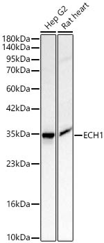
Western blot analysis of various lysates using ECH1 Rabbit pAb (CAB12944) at 1:2000 dilution. Secondary antibody: HRP-conjugated Goat anti-Rabbit IgG (H+L) (CABS014) at 1:10000 dilution. Lysates / proteins: 25 μg per lane. Blocking buffer: 3 % nonfat dry milk in TBST. Detection: ECL Basic Kit (AbGn00020). Exposure time: 30s.
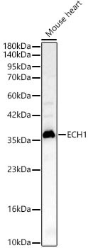
Western blot analysis of lysates from Mouse heart using ECH1 Rabbit pAb (CAB12944) at 1:2000 dilution. Secondary antibody: HRP-conjugated Goat anti-Rabbit IgG (H+L) (CABS014) at 1:10000 dilution. Lysates/proteins: 25 μg per lane. Blocking buffer: 3% nonfat dry milk in TBST. Detection: ECL Basic Kit (AbGn00020). Exposure time: 60s.