The ECSIT Antibody (CAB7804) is a high-quality antibody developed for reliable detection and analysis of target proteins. This antibody, generated in rabbits, demonstrates high reactivity with human samples and has been validated for Western blot applications.ECSIT is known for its role in the Toll-like receptor signaling pathway, which is crucial for the activation of the innate immune response. By targeting ECSIT, researchers can gain insights into the mechanisms underlying inflammatory signaling and immune system regulation. This antibody enables the detection and analysis of ECSIT in a variety of cell types, making it ideal for studies in immunology and infectious diseases.
This antibody is validated for use in WB, IF/ICC, ELISA applications and has demonstrated reactivity against Human, Mouse, Rat samples.
Product Name:
ECSIT Antibody
SKU:
CAB7804
Size:
20μL, 100μL
Reactivity:
Human, Mouse, Rat
Conjugate:
Unconjugated
Immunogen:
Recombinant protein (or fragment).This information is considered to be commercially sensitive.
Recommended starting concentration is 1 μg/mL. Please optimize the concentration based on your specific assay requirements.
Synonyms:
SITPEC, ECSIT
Positive Sample:
HepG2
Cellular Localization:
Cytoplasm, Mitochondrion, Nucleus.
Calculated MW:
49kDa
Observed MW:
49kDa
Predicted to enable DNA-binding transcription factor activity and chromatin binding activity. Involved in regulation of oxidoreductase activity and regulation of protein complex stability. Located in cytosol; mitochondrion; and nucleoplasm.
Purification Method
Affinity purification
Gene ID
51295
RRID
AB_2769260
Buffer Information
Store at -20℃. Avoid freeze / thaw cycles. Buffer: PBS containing 50% glycerol, preserved with proclin300 or sodium azide, pH 7.3.
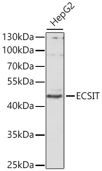
Western blot analysis of lysates from HepG2 cells, using ECSIT Rabbit pAb (CAB7804) at 1:1000 dilution. Secondary antibody: HRP-conjugated Goat anti-Rabbit IgG (H+L) (CABS014) at 1:10000 dilution. Lysates/proteins: 25μg per lane. Blocking buffer: 3% nonfat dry milk in TBST. Detection: ECL Basic Kit (AbGn00020). Exposure time: 30s.
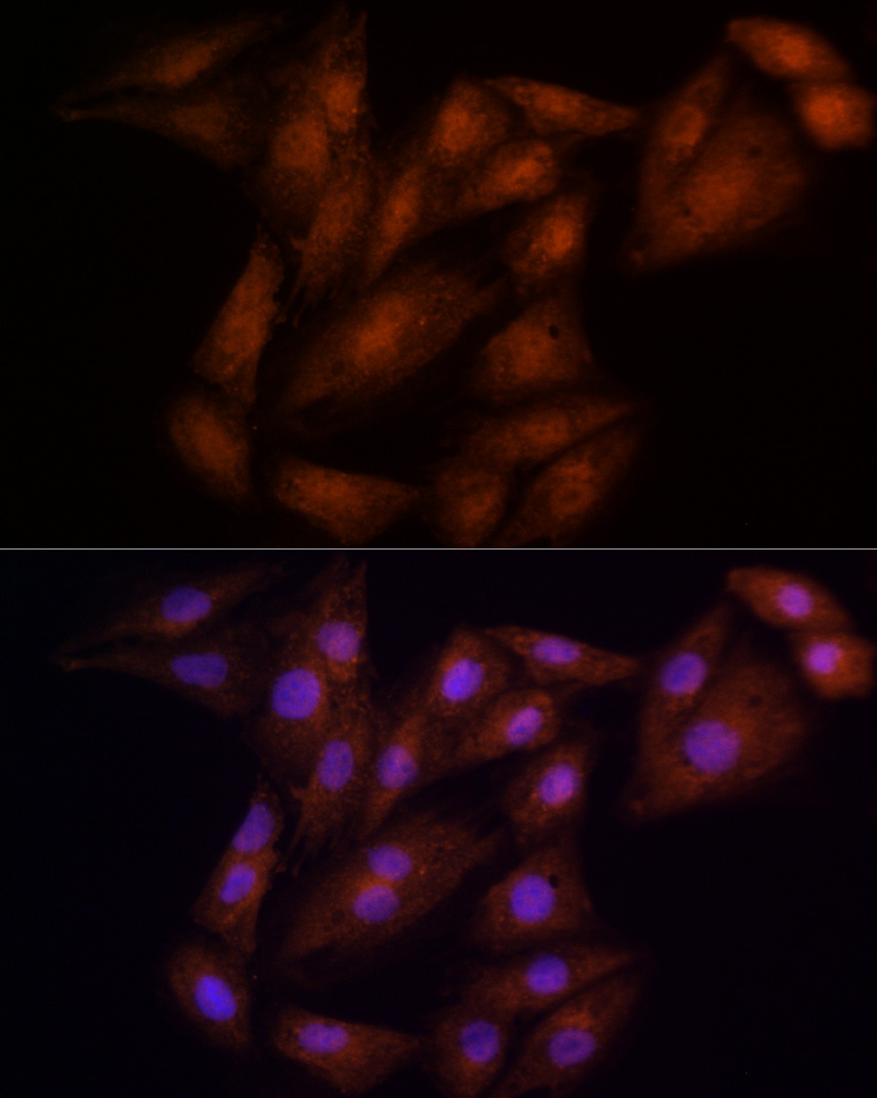
Immunofluorescence analysis of H9C2 cells using ECSIT Rabbit pAb (CAB7804) at dilution of 100 (40x lens). Secondary antibody: Cy3-conjugated Goat anti-Rabbit IgG (H+L) (CABS007) at 1:500 dilution. Blue: DAPI for nuclear staining.
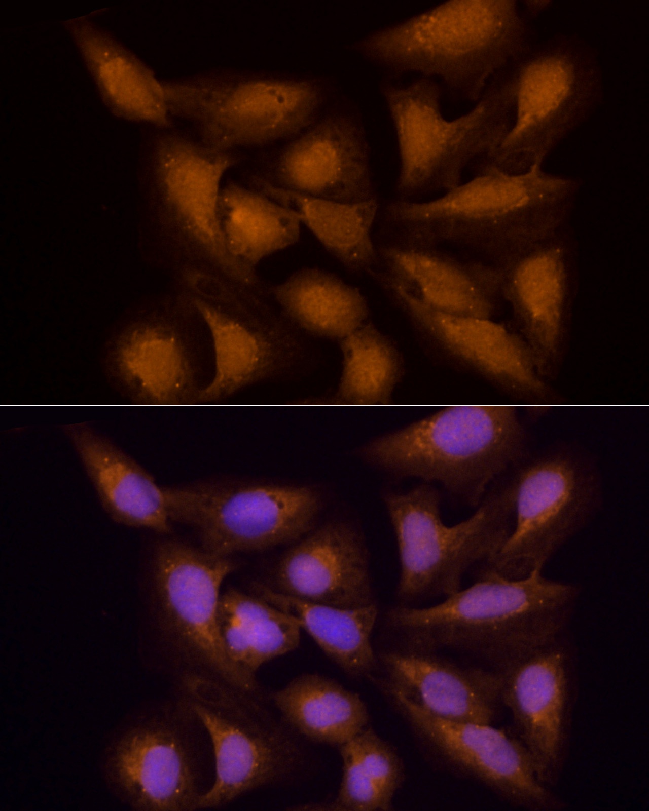
Immunofluorescence analysis of U2OS cells using ECSIT Rabbit pAb (CAB7804) at dilution of 100 (40x lens). Secondary antibody: Cy3-conjugated Goat anti-Rabbit IgG (H+L) (CABS007) at 1:500 dilution. Blue: DAPI for nuclear staining.