The EEF1D Antibody (CAB2509) is a high-quality antibody developed for reliable detection and analysis of target proteins. This antibody is produced in rabbits and has been extensively validated for use in Western blot applications, showing high reactivity with human samples. By specifically binding to EEF1D, this antibody allows for the accurate detection and analysis of the protein in various cell types.EEF1D, also known as elongation factor 1 delta, plays a crucial role in the elongation phase of protein synthesis and has been implicated in various cellular processes. Its role in translation regulation and signal transduction pathways make it a key player in cancer research, neurodegenerative diseases, and other disorders involving aberrant protein synthesis.
This antibody is validated for use in WB, IHC-P, IF/ICC, IP, ELISA applications and has demonstrated reactivity against Human, Mouse, Rat samples.
Product Name:
EEF1D Antibody
SKU:
CAB2509
Size:
20μL, 100μL
Reactivity:
Human, Mouse, Rat
Conjugate:
Unconjugated
Immunogen:
Recombinant protein (or fragment).This information is considered to be commercially sensitive.
0.5μg-4μg antibody for 200μg-400μg extracts of whole cells
ELISA
Recommended starting concentration is 1 μg/mL. Please optimize the concentration based on your specific assay requirements.
Synonyms:
EF1D, EF-1D, FP1047, EEF1D
Positive Sample:
MCF7, Mouse testis, jurkat, Jurkat
Cellular Localization:
Nucleus.
Calculated MW:
31kDa
Observed MW:
40kDa/35kDa/40kDa
This gene encodes a subunit of the elongation factor-1 complex, which is responsible for the enzymatic delivery of aminoacyl tRNAs to the ribosome. This subunit, delta, functions as guanine nucleotide exchange factor. It is reported that following HIV-1 infection, this subunit interacts with HIV-1 Tat. This interaction results in repression of translation of host cell proteins and enhanced translation of viral proteins. Several alternatively spliced transcript variants encoding multiple isoforms have been found for this gene. Related pseudogenes have been defined on chromosomes 1, 6, 7, 9, 11, 13, 17, 19.
Purification Method
Affinity purification
Gene ID
1936
RRID
AB_2764400
Buffer Information
Store at -20℃. Avoid freeze / thaw cycles. Buffer: PBS containing 50% glycerol, preserved with proclin300 or sodium azide, pH 7.3.
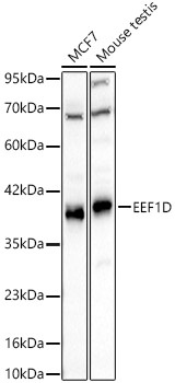
Western blot analysis of various lysates using EEF1D Rabbit pAb (CAB2509) at 1:2000 dilution. Secondary antibody: HRP-conjugated Goat anti-Rabbit IgG (H+L) (CABS014) at 1:10000 dilution. Lysates / proteins: 25 μg per lane. Blocking buffer: 3 % nonfat dry milk in TBST. Detection: ECL Basic Kit (AbGn00020). Exposure time: 10s.
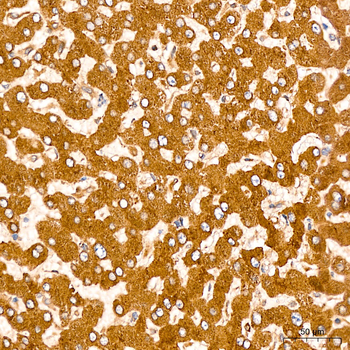
Immunohistochemistry analysis of paraffin-embedded Human liver tissue using EEF1D Rabbit pAb (CAB2509) at a dilution of 1:200 (40x lens). High pressure antigen retrieval was performed with 0.01 M citrate buffer (pH 6.0) prior to IHC staining.
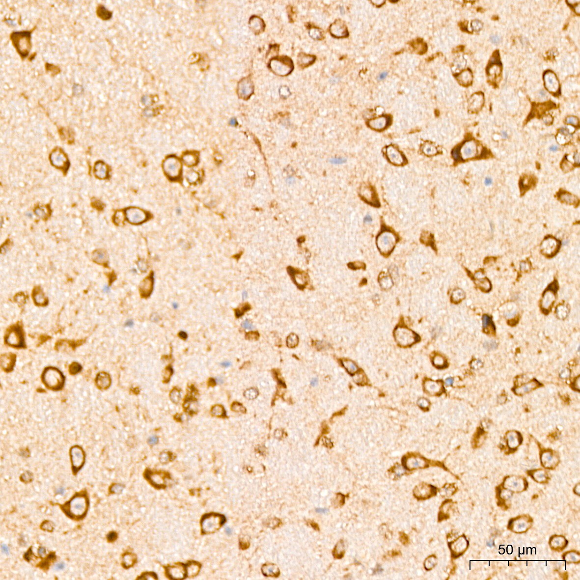
Immunohistochemistry analysis of paraffin-embedded Mouse brain tissue using EEF1D Rabbit pAb (CAB2509) at a dilution of 1:200 (40x lens). High pressure antigen retrieval was performed with 0.01 M citrate buffer (pH 6.0) prior to IHC staining.
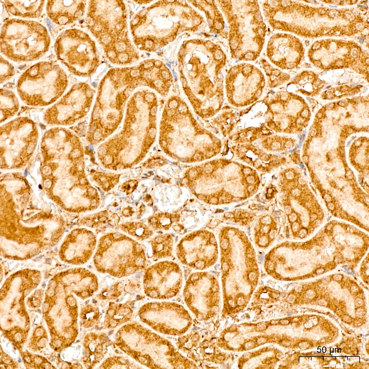
Immunohistochemistry analysis of paraffin-embedded Rat kidney tissue using EEF1D Rabbit pAb (CAB2509) at a dilution of 1:200 (40x lens). High pressure antigen retrieval was performed with 0.01 M citrate buffer (pH 6.0) prior to IHC staining.
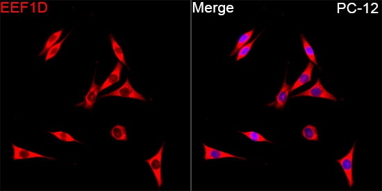
Immunofluorescence analysis of PC-12 cells using EEF1D Rabbit pAb (CAB2509) at dilution of 1:200 (40x lens). Secondary antibody: Cy3-conjugated Goat anti-Rabbit IgG (H+L) (CABS007) at 1:500 dilution. Blue: DAPI for nuclear staining.
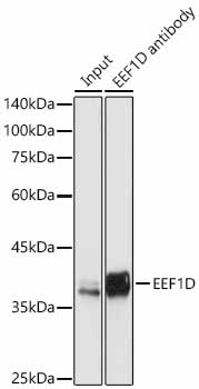
Immunoprecipitation of EEF1D in 300 µg extracts from jurkat cells using 3 µg EEF1D Rabbit pAb (CAB2509). Western blot analysis was performed using EEF1D Rabbit pAb (CAB2509) at 1:500 dilution.