The EFTUD2 Antibody (CAB7040) is a high-quality antibody developed for reliable detection and analysis of target proteins. This antibody, produced in rabbits, demonstrates high reactivity with human samples and has been validated for use in Western blot applications. By specifically binding to the EFTUD2 protein, this antibody enables precise detection and analysis in various cell types, making it an essential component for studies in molecular biology and genetic research.EFTUD2, also known as U5-116KD, plays a critical role in the spliceosome complex, which is essential for accurate RNA splicing and gene expression.
This antibody is validated for use in WB, IHC-P, IF/ICC, IP, ELISA applications and has demonstrated reactivity against Human, Mouse, Rat samples.
Product Name:
EFTUD2 Antibody
SKU:
CAB7040
Size:
20μL, 100μL
Reactivity:
Human, Mouse, Rat
Conjugate:
Unconjugated
Immunogen:
Recombinant protein (or fragment).This information is considered to be commercially sensitive.
This gene encodes a GTPase which is a component of the spliceosome complex which processes precursor mRNAs to produce mature mRNAs. Mutations in this gene are associated with mandibulofacial dysostosis with microcephaly. Multiple transcript variants encoding different isoforms have been found for this gene.
Purification Method
Affinity purification
Gene ID
9343
RRID
AB_2767595
Buffer Information
Store at -20℃. Avoid freeze / thaw cycles. Buffer: PBS containing 50% glycerol, preserved with proclin300 or sodium azide, pH 7.3.
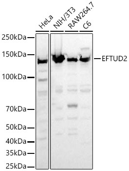
Western blot analysis of various lysates, using EFTUD2 Rabbit pAb (CAB7040) at 1:1000 dilution. Secondary antibody: HRP-conjugated Goat anti-Rabbit IgG (H+L) (CABS014) at 1:10000 dilution. Lysates/proteins: 25μg per lane. Blocking buffer: 3% nonfat dry milk in TBST. Detection: ECL Basic Kit (AbGn00020). Exposure time: 30s.
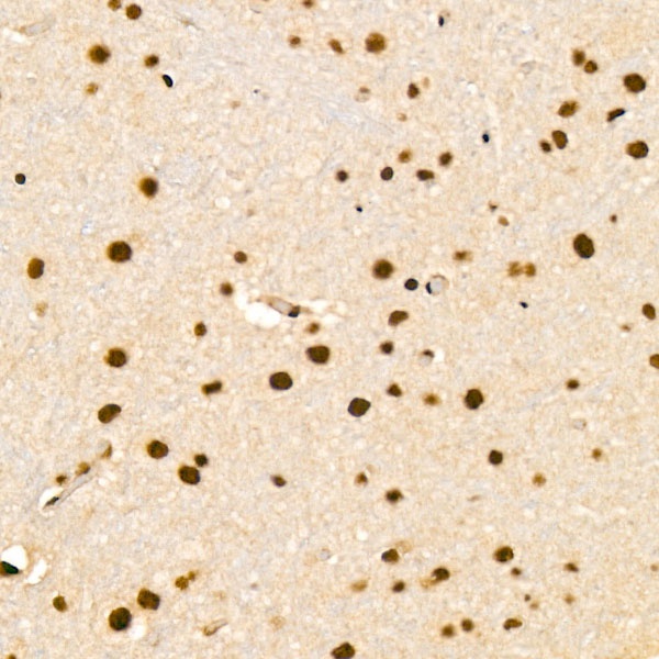
Immunohistochemistry analysis of paraffin-embedded Rat brain tissue using EFTUD2 Rabbit pAb (CAB7040) at a dilution of 1:100 (40x lens). High pressure antigen retrieval was performed with 0.01 M citrate buffer (pH 6.0) prior to IHC staining.
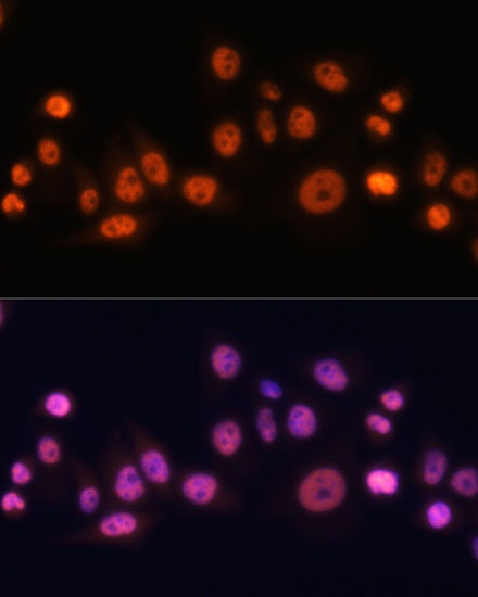
Immunofluorescence analysis of C6 cells using EFTUD2 Rabbit pAb (CAB7040) at dilution of 1:100. Secondary antibody: Cy3-conjugated Goat anti-Rabbit IgG (H+L) (CABS007) at 1:500 dilution. Blue: DAPI for nuclear staining.
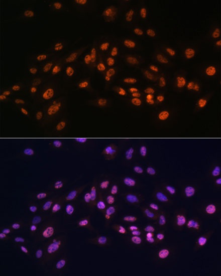
Immunofluorescence analysis of HeLa cells using EFTUD2 Rabbit pAb (CAB7040) at dilution of 1:100. Secondary antibody: Cy3-conjugated Goat anti-Rabbit IgG (H+L) (CABS007) at 1:500 dilution. Blue: DAPI for nuclear staining.
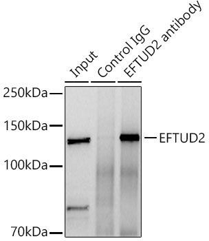
Immunoprecipitation analysis of 300 μg extracts of NTH-3T3 cells using 3 μg EFTUD2 Rabbit pAb (CAB7040). Western blot was performed from the immunoprecipitate using EFTUD2 Rabbit pAb (CAB7040) at a dilition of 1:2000.