The EGFL7 Antibody (CAB9376) is a high-quality antibody developed for reliable detection and analysis of target proteins. This gene encodes a secreted endothelial cell protein that contains two epidermal growth factor-like domains. The encoded protein may play a role in regulating vasculogenesis. This protein may be involved in the growth and proliferation of tumor cells. Alternate splicing results in multiple transcript variants.
This antibody is validated for use in WB, ELISA applications and has demonstrated reactivity against Mouse, Rat samples.
Product Name:
EGFL7 Antibody
SKU:
CAB9376
Size:
100μL, 20μL
Reactivity:
Mouse, Rat
Conjugate:
Unconjugated
Immunogen:
Recombinant protein (or fragment).This information is considered to be commercially sensitive.
Tested Applications:
WBELISA
Recommended Dilution:
WB
1:500 - 1:2000
ELISA
Recommended starting concentration is 1 μg/mL. Please optimize the concentration based on your specific assay requirements.
Synonyms:
NEU1, ZNEU1, VE-STATIN, EGFL7
Positive Sample:
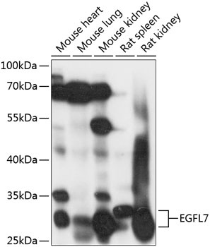
Mouse heart, Mouse lung, Mouse kidney, Rat spleen, Rat kidney
Cellular Localization:
Secreted, Extracellular Space.
Calculated MW:
30kDa
Observed MW:
30kDa
This gene encodes a secreted endothelial cell protein that contains two epidermal growth factor-like domains. The encoded protein may play a role in regulating vasculogenesis. This protein may be involved in the growth and proliferation of tumor cells. Alternate splicing results in multiple transcript variants.
Purification Method
Affinity purification
Gene ID
51162
RRID
AB_2769275
Buffer Information
Store at -20℃. Avoid freeze / thaw cycles. Buffer: PBS with 0.01% thimerosal,50% glycerol,pH7.3.
Western blot analysis of various lysates using EGFL7 Rabbit pAb (CAB9376) at 1:1000 dilution. Secondary antibody: HRP-conjugated Goat anti-Rabbit IgG (H+L) (AS014) at 1:10000 dilution. Lysates/proteins: 25μg per lane. Blocking buffer: 3% nonfat dry milk in TBST. Detection: ECL Basic Kit (AbGn00020). Exposure time: 10s.