The PKR/EIF2AK2 Monoclonal Antibody (CAB19545) is a high-quality antibody developed for reliable detection and analysis of target proteins. This monoclonal antibody, derived from rabbit sources, is highly specific and sensitive for detecting PKR in various experimental settings.PKR is a serine/threonine protein kinase that is activated in response to viral infection, leading to the inhibition of protein synthesis and the induction of apoptosis in infected cells. By targeting PKR with this antibody, researchers can study its role in antiviral defense mechanisms, as well as its involvement in other cellular processes such as inflammation and stress response.
This antibody is validated for use in WB, IF/ICC, ELISA applications and has demonstrated reactivity against Human samples.
Product Name:
PKR/EIF2AK2 Monoclonal Antibody
SKU:
CAB19545
Size:
20μL, 100μL
Reactivity:
Human
Clone Number:
ARC0024
Conjugate:
Unconjugated
Immunogen:
Synthetic peptide. This information is considered to be commercially sensitive.
The protein encoded by this gene is a serine/threonine protein kinase that is activated by autophosphorylation after binding to dsRNA. The activated form of the encoded protein can phosphorylate translation initiation factor EIF2S1, which in turn inhibits protein synthesis. This protein is also activated by manganese ions and heparin. The encoded protein plays an important role in the innate immune response against multiple DNA and RNA viruses.
Purification Method
Affinity purification
Gene ID
5610
RRID
AB_2862660
Buffer Information
Store at -20℃. Avoid freeze / thaw cycles. Buffer: PBS containing 50% glycerol and 0.05% BSA, preserved with proclin300 or sodium azide, pH 7.3.
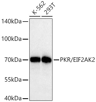
Western blot analysis of various lysates using PKR/EIF2AK2 Rabbit mAb (CAB19545) at 1:2000 dilution incubated overnight at 4℃. Secondary antibody: HRP-conjugated Goat anti-Rabbit IgG (H+L) (CABS014) at 1:10000 dilution. Lysates/proteins: 25 μg per lane. Blocking buffer: 3% nonfat dry milk in TBST. Detection: ECL Basic Kit (AbGn00020). Exposure time: 30 s.
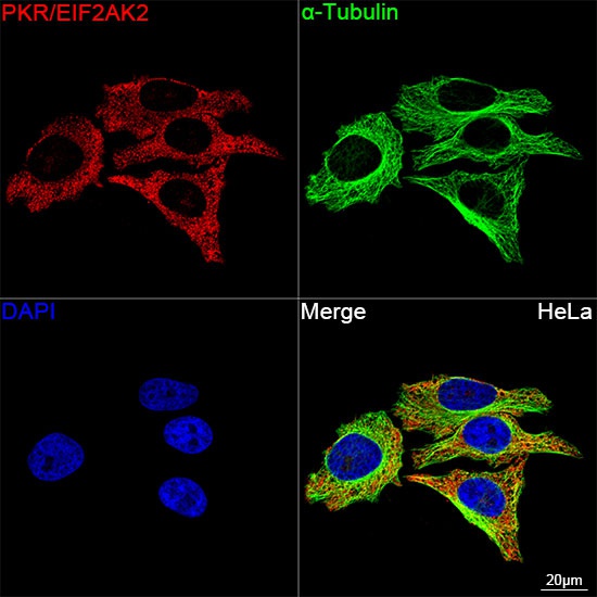
Confocal imaging of HeLa cells using PKR/EIF2AK2 Rabbit mAb (CAB19545, dilution 1:50) followed by a further incubation with Cy3 Goat Anti-Rabbit IgG (H+L) (CABS007, dilution 1:500) (Red). The cells were counterstained with α-Tubulin Mouse mAb (AC012, dilution 1:400) followed by incubation with ABflo® 488-conjugated Goat Anti-Mouse IgG (H+L) Ab (CABS076, dilution 1:500) (Green). DAPI was used for nuclear staining (Blue). Objective: 100x.


")
")