The EIF3D Antibody (CAB5947) is a high-quality antibody developed for reliable detection and analysis of target proteins. This antibody, produced in rabbits, exhibits high reactivity with human samples and is specifically validated for use in Western blot applications. With its ability to bind to the EIF3D protein, researchers can accurately detect and analyze EIF3D expression in a variety of cell types, making it an essential tool for studies in molecular biology and translational research.EIF3D plays a crucial role in the initiation of protein translation, making it a key player in cellular processes such as cell growth, differentiation, and proliferation.
This antibody is validated for use in WB, IF/ICC, ELISA applications and has demonstrated reactivity against Human, Mouse, Rat samples.
Product Name:
EIF3D Antibody
SKU:
CAB5947
Size:
20μL, 100μL
Reactivity:
Human, Mouse, Rat
Conjugate:
Unconjugated
Immunogen:
Recombinant protein (or fragment).This information is considered to be commercially sensitive.
Recommended starting concentration is 1 μg/mL. Please optimize the concentration based on your specific assay requirements.
Synonyms:
EIF3S7, eIF3-p66, eIF3-zeta, EIF3D
Positive Sample:
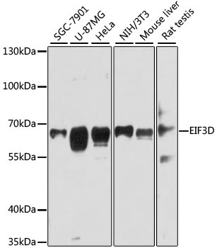
SGC-7901, U-87MG, HeLa, NIH/3T3, Mouse liver, Rat testis
Cellular Localization:
Cytoplasm.
Calculated MW:
64kDa
Observed MW:
64kDa
Eukaryotic translation initiation factor-3 (eIF3), the largest of the eIFs, is a multiprotein complex composed of at least ten nonidentical subunits. The complex binds to the 40S ribosome and helps maintain the 40S and 60S ribosomal subunits in a dissociated state. It is also thought to play a role in the formation of the 40S initiation complex by interacting with the ternary complex of eIF2/GTP/methionyl-tRNA, and by promoting mRNA binding. The protein encoded by this gene is the major RNA binding subunit of the eIF3 complex.
Purification Method
Affinity purification
Gene ID
8664
RRID
AB_2766679
Buffer Information
Store at -20℃. Avoid freeze / thaw cycles. Buffer: PBS with 0.01% thimerosal,50% glycerol,pH7.3.
Western blot analysis of various lysates using EIF3D Rabbit pAb (CAB5947) at 1:3000 dilution. Secondary antibody: HRP-conjugated Goat anti-Rabbit IgG (H+L) (CABS014) at 1:10000 dilution. Lysates/proteins: 25μg per lane. Blocking buffer: 3% nonfat dry milk in TBST. Detection: ECL Basic Kit (AbGn00020). Exposure time: 30s.
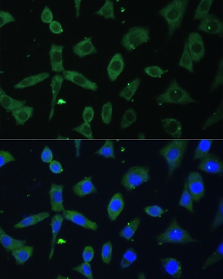
Immunofluorescence analysis of L-929 cells using EIF3D Rabbit pAb (CAB5947) at dilution of 1:100. Secondary antibody: Cy3-conjugated Goat anti-Rabbit IgG (H+L) (CABS007) at 1:500 dilution. Blue: DAPI for nuclear staining.
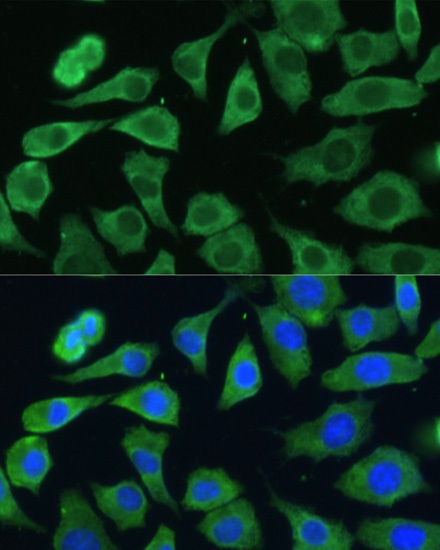
Immunofluorescence analysis of L-929 cells using EIF3D Rabbit pAb (CAB5947) at dilution of 1:100. Secondary antibody: Cy3-conjugated Goat anti-Rabbit IgG (H+L) (CABS007) at 1:500 dilution. Blue: DAPI for nuclear staining.
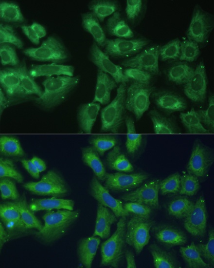
Immunofluorescence analysis of U-2 OS cells using EIF3D Rabbit pAb (CAB5947) at dilution of 1:100. Secondary antibody: Cy3-conjugated Goat anti-Rabbit IgG (H+L) (CABS007) at 1:500 dilution. Blue: DAPI for nuclear staining.