The EIF3F Antibody (CAB7023) is a high-quality antibody developed for reliable detection and analysis of target proteins. This antibody, produced in rabbits, has high specificity and sensitivity for detecting EIF3F in human samples, making it ideal for use in Western blot applications.EIF3F plays a crucial role in the initiation of protein synthesis, making it a vital component in cellular processes such as cell growth and proliferation. Dysfunction of EIF3F has been implicated in various diseases, including cancer, making it an important target for research in oncology and molecular biology.
This antibody is validated for use in WB, IHC-P, IF/ICC, IP, ELISA applications and has demonstrated reactivity against Human, Mouse, Rat samples.
Product Name:
EIF3F Antibody
SKU:
CAB7023
Size:
20μL, 100μL
Reactivity:
Human, Mouse, Rat
Conjugate:
Unconjugated
Immunogen:
Recombinant protein (or fragment).This information is considered to be commercially sensitive.
0.5μg-4μg antibody for 200μg-400μg extracts of whole cells
ELISA
Recommended starting concentration is 1 μg/mL. Please optimize the concentration based on your specific assay requirements.
Synonyms:
MRT67, EIF3S5, eIF3-p47, EIF3F
Positive Sample:
HeLa
Cellular Localization:
Cytoplasm.
Calculated MW:
38kDa
Observed MW:
38kDa
Enables deubiquitinase activity and identical protein binding activity. Contributes to translation initiation factor activity. Involved in IRES-dependent viral translational initiation; protein deubiquitination; and translational initiation. Located in membrane. Part of eukaryotic translation initiation factor 3 complex. Implicated in autosomal recessive non-syndromic intellectual disability.
Purification Method
Affinity purification
Gene ID
8665
RRID
AB_2767579
Buffer Information
Store at -20℃. Avoid freeze / thaw cycles. Buffer: PBS containing 50% glycerol, preserved with proclin300 or sodium azide, pH 7.3.
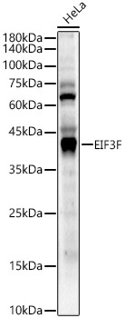
Western blot analysis of lysates from HeLa cells, using EIF3F Rabbit pAb (CAB7023) at 1:1000 dilution. Secondary antibody: HRP-conjugated Goat anti-Rabbit IgG (H+L) (CABS014) at 1:10000 dilution. Lysates/proteins: 25μg per lane. Blocking buffer: 3% nonfat dry milk in TBST. Detection: ECL Enhanced Kit (AbGn00021). Exposure time: 60s.
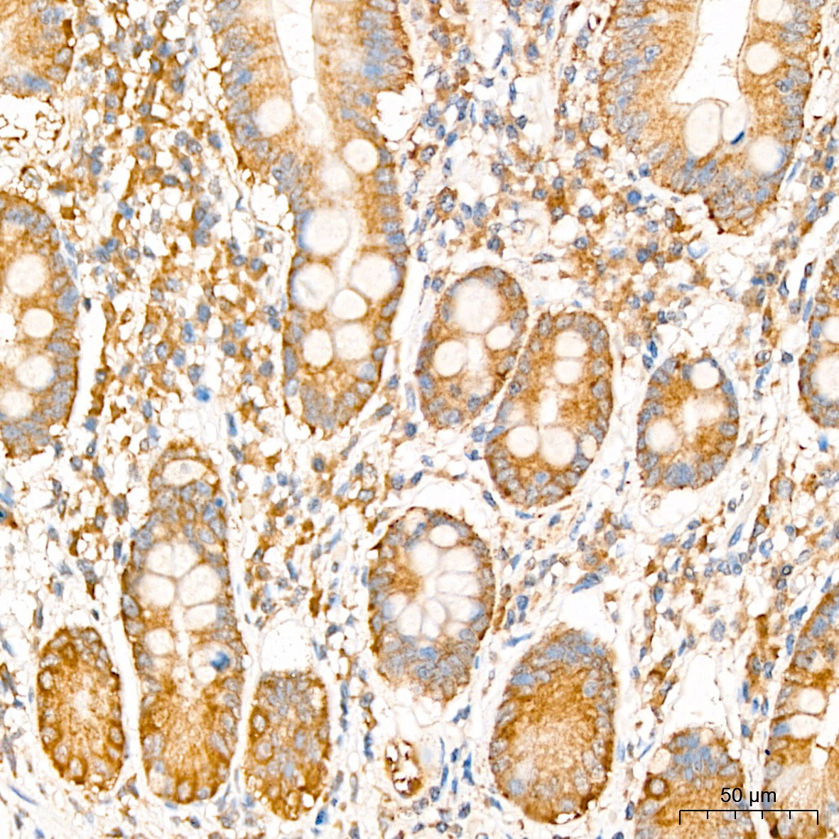
Immunohistochemistry analysis of paraffin-embedded Human colon tissue using EIF3F Rabbit pAb (CAB7023) at a dilution of 1:200 (40x lens). High pressure antigen retrieval was performed with 0.01 M citrate buffer (pH 6.0) prior to IHC staining.
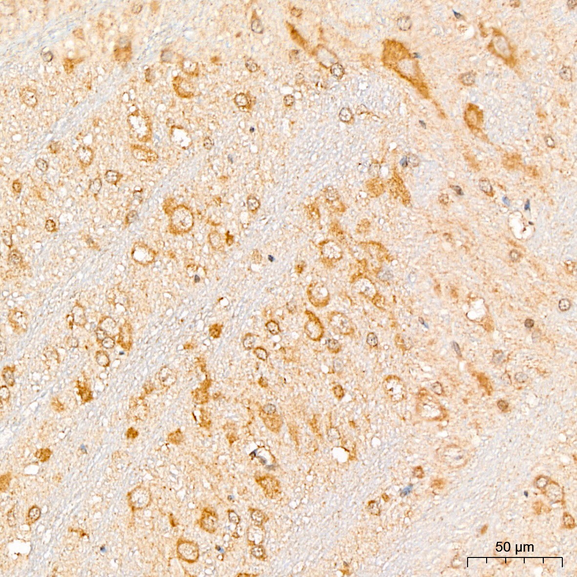
Immunohistochemistry analysis of paraffin-embedded Rat brain tissue using EIF3F Rabbit pAb (CAB7023) at a dilution of 1:200 (40x lens). High pressure antigen retrieval was performed with 0.01 M citrate buffer (pH 6.0) prior to IHC staining.
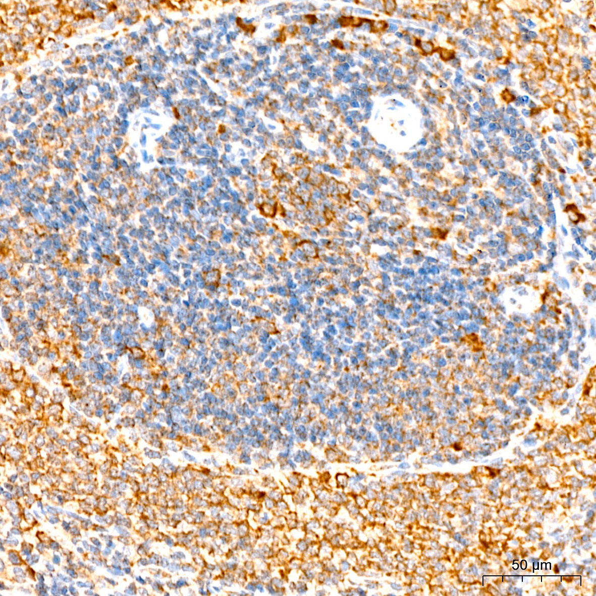
Immunohistochemistry analysis of paraffin-embedded Mouse spleen tissue using EIF3F Rabbit pAb (CAB7023) at a dilution of 1:200 (40x lens). High pressure antigen retrieval was performed with 0.01 M citrate buffer (pH 6.0) prior to IHC staining.
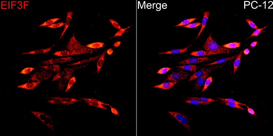
Immunofluorescence analysis of PC-12 cells using EIF3F Rabbit pAb (CAB7023) at dilution of 1:200 (40x lens). Secondary antibody: Cy3-conjugated Goat anti-Rabbit IgG (H+L) (CABS007) at 1:500 dilution. Blue: DAPI for nuclear staining.
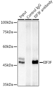
Immunoprecipitation analysis of 300 μg extracts of Jurkat cells using 3 μg EIF3F antibody (CAB7023). Western blot was performed from the immunoprecipitate using EIF3F antibody (CAB7023) at a dilution of 1:1000.