The EIF3F Antibody (CAB7023) is a high-quality antibody developed for reliable detection and analysis of target proteins. Enables deubiquitinase activity and identical protein binding activity. Contributes to translation initiation factor activity. Involved in IRES-dependent viral translational initiation; protein deubiquitination; and translational initiation. Located in membrane. Part of eukaryotic translation initiation factor 3 complex. Implicated in autosomal recessive non-syndromic intellectual disability.
This antibody is validated for use in WB, IHC-P, IF/ICC, IP, ELISA applications and has demonstrated reactivity against Human, Mouse, Rat samples.
Product Name:
EIF3F Antibody
SKU:
CAB7023
Size:
100μL, 20μL
Reactivity:
Human, Mouse, Rat
Conjugate:
Unconjugated
Immunogen:
Recombinant protein (or fragment).This information is considered to be commercially sensitive.
Tested Applications:
WBIHC-PIF/ICCIPELISA
Recommended Dilution:
WB
1:500 - 1:1000
IHC-P
1:50 - 1:200
IF/ICC
1:50 - 1:200
IP
0.5μg-4μg antibody for 200μg-400μg extracts of whole cells
ELISA
Recommended starting concentration is 1 μg/mL. Please optimize the concentration based on your specific assay requirements.
Synonyms:
MRT67, EIF3S5, eIF3-p47, EIF3F
Positive Sample:
HeLa
Cellular Localization:
Cytoplasm.
Calculated MW:
38kDa
Observed MW:
38kDa
Enables deubiquitinase activity and identical protein binding activity. Contributes to translation initiation factor activity. Involved in IRES-dependent viral translational initiation; protein deubiquitination; and translational initiation. Located in membrane. Part of eukaryotic translation initiation factor 3 complex. Implicated in autosomal recessive non-syndromic intellectual disability.
Purification Method
Affinity purification
Gene ID
8665
RRID
AB_2767579
Buffer Information
Store at -20℃. Avoid freeze / thaw cycles. Buffer: PBS containing 50% glycerol, preserved with proclin300 or sodium azide, pH 7.3.
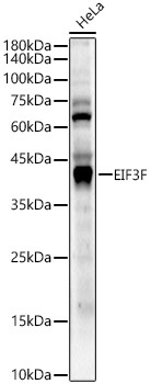
Western blot analysis of lysates from HeLa cells, using EIF3F Rabbit pAb (CAB7023) at 1:1000 dilution. Secondary antibody: HRP-conjugated Goat anti-Rabbit IgG (H+L) (AS014) at 1:10000 dilution. Lysates/proteins: 25μg per lane. Blocking buffer: 3% nonfat dry milk in TBST. Detection: ECL Enhanced Kit (AbGn00021). Exposure time: 60s.
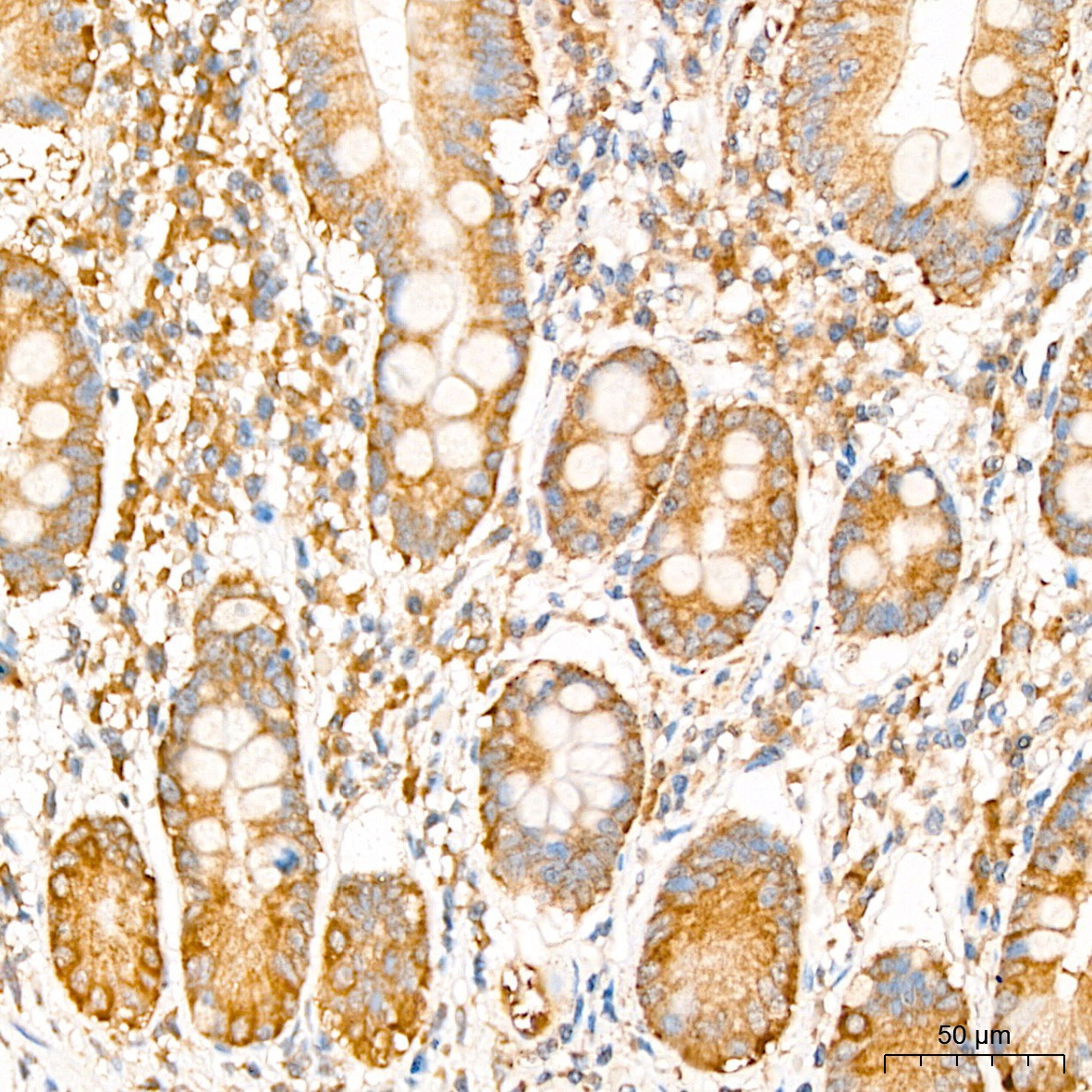
Immunohistochemistry analysis of paraffin-embedded Human colon tissue using EIF3F Rabbit pAb (CAB7023) at a dilution of 1:200 (40x lens). High pressure antigen retrieval was performed with 0.01 M citrate buffer (pH 6.0) prior to IHC staining.
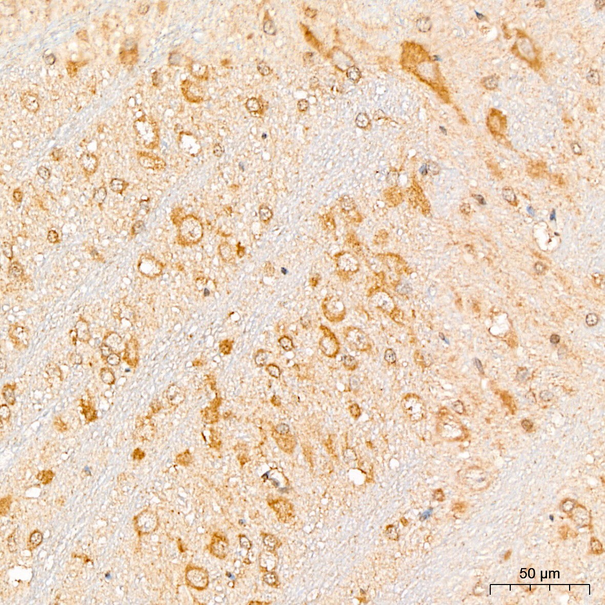
Immunohistochemistry analysis of paraffin-embedded Rat brain tissue using EIF3F Rabbit pAb (CAB7023) at a dilution of 1:200 (40x lens). High pressure antigen retrieval was performed with 0.01 M citrate buffer (pH 6.0) prior to IHC staining.
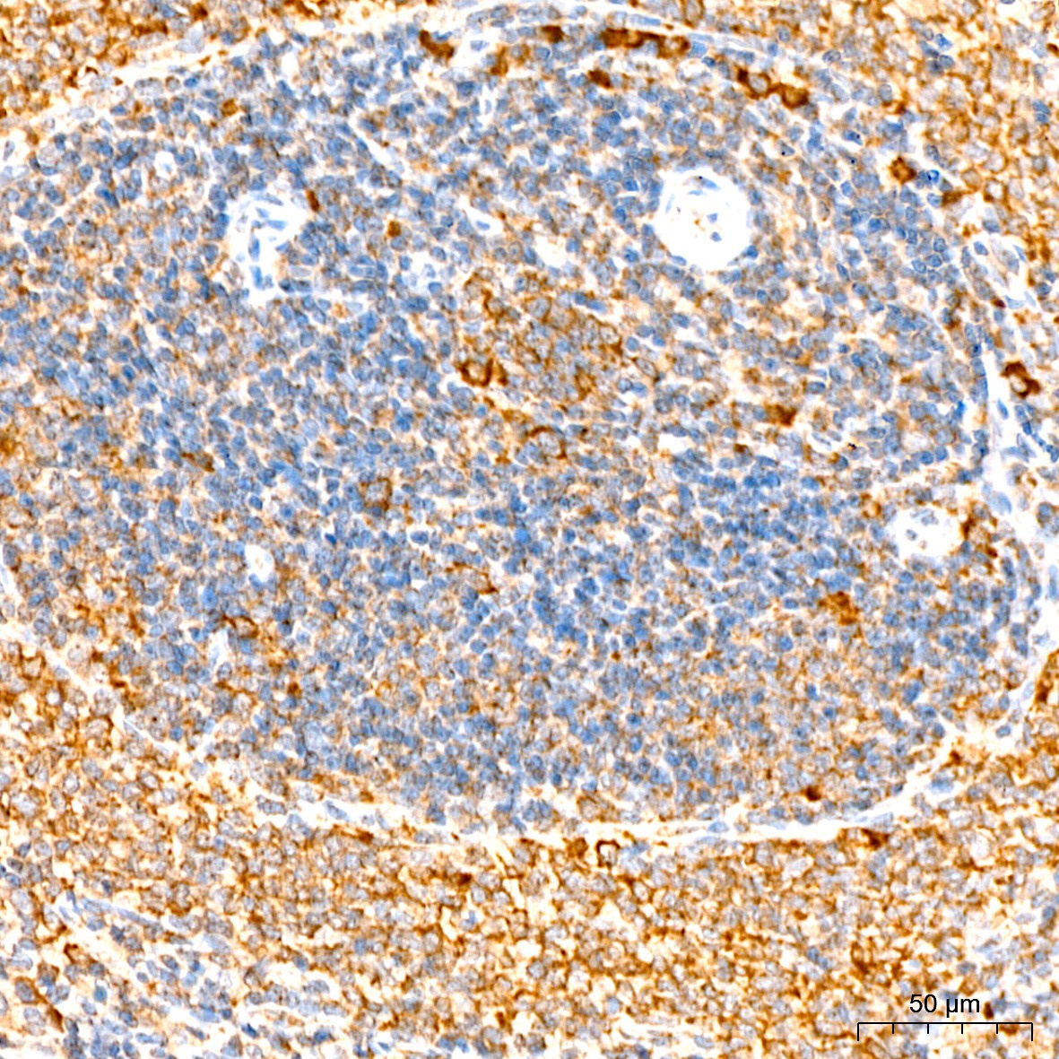
Immunohistochemistry analysis of paraffin-embedded Mouse spleen tissue using EIF3F Rabbit pAb (CAB7023) at a dilution of 1:200 (40x lens). High pressure antigen retrieval was performed with 0.01 M citrate buffer (pH 6.0) prior to IHC staining.
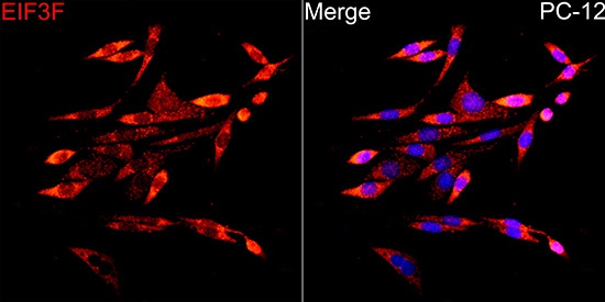
Immunofluorescence analysis of PC-12 cells using EIF3F Rabbit pAb (CAB7023) at dilution of 1:200 (40x lens). Secondary antibody: Cy3-conjugated Goat anti-Rabbit IgG (H+L) (AS007) at 1:500 dilution. Blue: DAPI for nuclear staining.
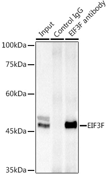
Immunoprecipitation analysis of 300 μg extracts of Jurkat cells using 3 μg EIF3F antibody (CAB7023). Western blot was performed from the immunoprecipitate using EIF3F antibody (CAB7023) at a dilution of 1:1000.