The EIF3M Antibody (CAB4426) is a high-quality antibody developed for reliable detection and analysis of target proteins. This antibody, produced in rabbits, is highly specific to human EIF3M and has been validated for use in Western blot applications.EIF3M is essential for protein synthesis and plays a crucial role in the regulation of translation initiation. Dysregulation of EIF3M has been linked to various diseases, including cancer, making it a promising target for therapeutic interventions. By detecting and analyzing EIF3M expression in different cell types, this antibody is ideal for studies focusing on protein synthesis, gene expression, and molecular mechanisms involved in disease pathogenesis.
This antibody is validated for use in WB, IHC-P, ELISA applications and has demonstrated reactivity against Human, Mouse, Rat samples.
Product Name:
EIF3M Antibody
SKU:
CAB4426
Size:
20μL, 100μL
Reactivity:
Human, Mouse, Rat
Conjugate:
Unconjugated
Immunogen:
Recombinant protein (or fragment).This information is considered to be commercially sensitive.
Recommended starting concentration is 1 μg/mL. Please optimize the concentration based on your specific assay requirements.
Synonyms:
B5, GA17, PCID1, TANGO7, hfl-B5, EIF3M
Positive Sample:
HeLa, A-549, HepG2, NCI-H460, Mouse spleen, Mouse heart, Mouse thymus, Rat spleen, Rat thymus, HeLa, Mouse thymus, Rat thymus
Cellular Localization:
Cytoplasm.
Calculated MW:
42kDa
Observed MW:
40kDa/36kDa
This gene encodes a protein that is part of the eurkaryotic translation initiation factor 3 complete (eIF-3) required for protein synthesis. Elevated levels of the encoded protein are present in cancer cell lines. Inactivation of the encoded protein has been shown to interfere with translation of herpes virus mRNAs by preventing the association of mRNAs with the ribosomes. A pseudogene of this gene is located on the X chromosome.
Purification Method
Affinity purification
Gene ID
10480
RRID
AB_2765669
Buffer Information
Store at -20℃. Avoid freeze / thaw cycles. Buffer: PBS containing 50% glycerol, preserved with proclin300 or sodium azide, pH 7.3.
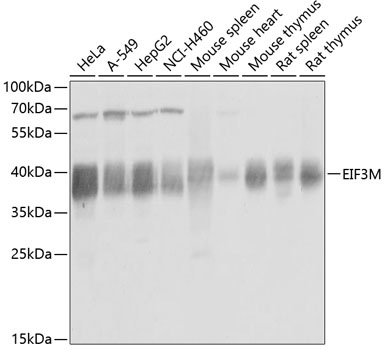
Western blot analysis of various lysates using EIF3M Rabbit pAb (CAB4426) at 1:1000 dilution. Secondary antibody: HRP-conjugated Goat anti-Rabbit IgG (H+L) (CABS014) at 1:10000 dilution. Lysates/proteins: 25μg per lane. Blocking buffer: 3% nonfat dry milk in TBST. Detection: ECL Basic Kit (AbGn00020). Exposure time: 1s.
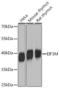
Western blot analysis of various lysates using EIF3M Rabbit pAb (CAB4426) at 1:1000 dilution incubated overnight at 4℃. Secondary antibody: HRP-conjugated Goat anti-Rabbit IgG (H+L) (CABS014) at 1:10000 dilution. Lysates/proteins: 25 μg per lane. Blocking buffer: 3% nonfat dry milk in TBST. Detection: ECL Basic Kit (AbGn00020) Exposure time: 5 s.
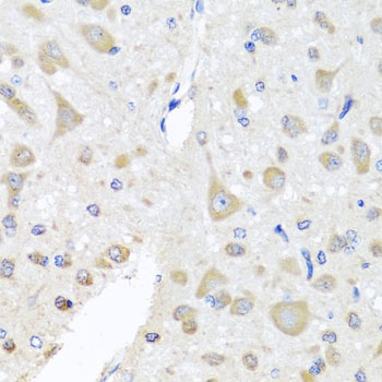
Immunohistochemistry analysis of paraffin-embedded Rat brain using EIF3M Rabbit pAb (CAB4426) at dilution of 1:100 (40x lens). Microwave antigen retrieval performed with 0.01M PBS Buffer (pH 7.2) prior to IHC staining.