The EML4 Antibody (CAB16117) is a high-quality antibody developed for reliable detection and analysis of target proteins. This antibody, produced in rabbits, is highly specific to human samples and has been validated for use in Western blot applications.EML4, or echinoderm microtubule-associated protein-like 4, is a key player in the development and progression of certain cancer types. Its fusion with other genes can lead to the activation of oncogenic pathways, making it a promising target for therapeutic interventions.
This antibody is validated for use in WB, IHC-P, IF/ICC, ELISA applications and has demonstrated reactivity against Human, Mouse, Rat samples.
Product Name:
EML4 Antibody
SKU:
CAB16117
Size:
20μL, 100μL
Reactivity:
Human, Mouse, Rat
Conjugate:
Unconjugated
Immunogen:
Recombinant protein (or fragment).This information is considered to be commercially sensitive.
Recommended starting concentration is 1 μg/mL. Please optimize the concentration based on your specific assay requirements.
Synonyms:
C2orf2, ELP120, EMAP-4, EMAPL4, ROPP120, EML4
Positive Sample:
HepG2
Cellular Localization:
Cytoplasm, Cytoskeleton.
Calculated MW:
109kDa
Observed MW:
120kDa
This gene is a member of the echinoderm microtubule associated protein-like family. The encoded WD-repeat protein may be involved in microtubule formation. Abnormal fusion of parts of this gene with portions of the anaplastic lymphoma receptor tyrosine kinase gene, which generates EML4-ALK fusion transcripts, is one of the primary mutations associated with non-small cell lung cancer. Alternative splicing of this gene results in two transcript variants.
Purification Method
Affinity purification
Gene ID
27436
RRID
AB_2763561
Buffer Information
Store at -20℃. Avoid freeze / thaw cycles. Buffer: PBS with 0.01% thimerosal,50% glycerol,pH7.3.
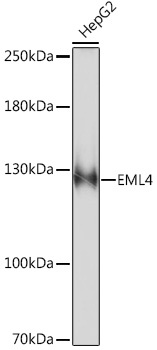
Western blot analysis of lysates from HepG2 cells, using EML4 Rabbit pAb (CAB16117) at 1:1000 dilution. Secondary antibody: HRP-conjugated Goat anti-Rabbit IgG (H+L) (CABS014) at 1:10000 dilution. Lysates/proteins: 25μg per lane. Blocking buffer: 3% nonfat dry milk in TBST. Detection: ECL Basic Kit (AbGn00020). Exposure time: 90s.
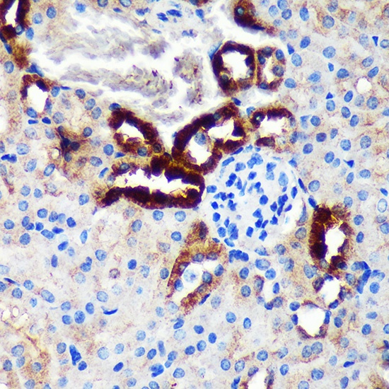
Immunohistochemistry analysis of paraffin-embedded Mouse kidney using EML4 Rabbit pAb (CAB16117) at dilution of 1:100 (40x lens). Microwave antigen retrieval performed with 0.01M PBS Buffer (pH 7.2) prior to IHC staining.
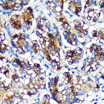
Immunohistochemistry analysis of paraffin-embedded Human liver cancer using EML4 Rabbit pAb (CAB16117) at dilution of 1:100 (40x lens). Microwave antigen retrieval performed with 0.01M PBS Buffer (pH 7.2) prior to IHC staining.
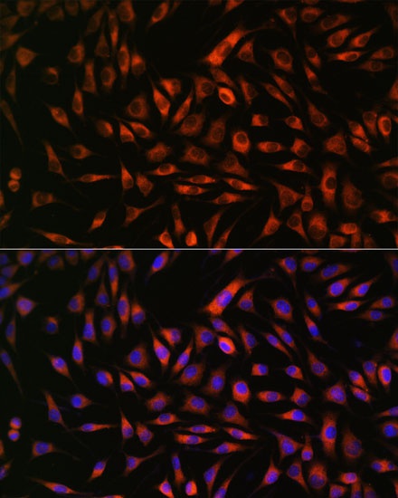
Immunofluorescence analysis of L929 cells using EML4 Rabbit pAb (CAB16117) at dilution of 1:100. Secondary antibody: Cy3-conjugated Goat anti-Rabbit IgG (H+L) (CABS007) at 1:500 dilution. Blue: DAPI for nuclear staining.