The SH3GL1 Antibody (CAB7449) is a high-quality antibody developed for reliable detection and analysis of target proteins. This antibody, produced in rabbits, exhibits high reactivity with human samples and has been validated for use in Western blot applications.Endophilin A2 is a protein involved in the formation of vesicles that transport molecules within cells, making it essential for various cellular processes including synaptic vesicle recycling and protein trafficking. Research on Endophilin A2 is important for understanding how cells regulate their internal environment and communicate with other cells through vesicle transport mechanisms.
This antibody is validated for use in WB, ELISA applications and has demonstrated reactivity against Human, Mouse, Rat, Monkey samples.
Product Name:
SH3GL1 Antibody
SKU:
CAB7449
Size:
20μL, 100μL
Reactivity:
Human, Mouse, Rat, Monkey
Conjugate:
Unconjugated
Immunogen:
Recombinant protein (or fragment).This information is considered to be commercially sensitive.
Recommended starting concentration is 1 μg/mL. Please optimize the concentration based on your specific assay requirements.
Synonyms:
EEN, CNSA1, SH3P8, SH3D2B, SH3GL1
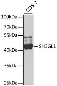
Positive Sample:
COS-7
Cellular Localization:
Cell Projection, Cytoplasm, Early Endosome Membrane, Peripheral Membrane Protein, Podosome.
Calculated MW:
41kDa
Observed MW:
41kDa
This gene encodes a member of the endophilin family of Src homology 3 domain-containing proteins. The encoded protein is involved in endocytosis and may also play a role in the cell cycle. Overexpression of this gene may play a role in leukemogenesis, and the encoded protein has been implicated in acute myeloid leukemia as a fusion partner of the myeloid-lymphoid leukemia protein. Pseudogenes of this gene are located on the long arm of chromosomes 11 and 17. Alternatively spliced transcript variants encoding multiple isoforms have been observed for this gene.
Purification Method
Affinity purification
Gene ID
6455
RRID
AB_2767980
Buffer Information
Store at -20℃. Avoid freeze / thaw cycles. Buffer: PBS containing 50% glycerol, preserved with proclin300 or sodium azide, pH 7.3.
Western blot analysis of lysates from COS-7 cells, using SH3GL1 Rabbit pAb (CAB7449) at 1:1000 dilution. Secondary antibody: HRP-conjugated Goat anti-Rabbit IgG (H+L) (CABS014) at 1:10000 dilution. Lysates/proteins: 25μg per lane. Blocking buffer: 3% nonfat dry milk in TBST. Detection: ECL Basic Kit (AbGn00020). Exposure time: 60s.