The EPHA5 Antibody (CAB14238) is a high-quality antibody developed for reliable detection and analysis of target proteins. This antibody, produced in rabbits, exhibits high reactivity with human samples and has been rigorously validated for use in Western blot applications.EPHA5 is known to play a key role in various biological processes, including cell adhesion, migration, and axon guidance. Dysregulation of EPHA5 has been implicated in neurological disorders, cancer, and other diseases, making it an attractive target for further research.
This antibody is validated for use in WB, IF/ICC, ELISA applications and has demonstrated reactivity against Human, Mouse, Rat samples.
Product Name:
EPHA5 Antibody
SKU:
CAB14238
Size:
20μL, 100μL
Reactivity:
Human, Mouse, Rat
Conjugate:
Unconjugated
Immunogen:
Recombinant protein (or fragment).This information is considered to be commercially sensitive.
Recommended starting concentration is 1 μg/mL. Please optimize the concentration based on your specific assay requirements.
Synonyms:
EK7, CEK7, EHK1, HEK7, EHK-1, TYRO4, EPHA5
Positive Sample:
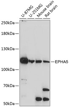
U-87MG, U-251MG, Mouse brain, Rat brain
Cellular Localization:
Cell Membrane, Cell Projection, Single-Pass Type I Membrane Protein, Axon, Dendrite.
Calculated MW:
115kDa
Observed MW:
115kDa
This gene belongs to the ephrin receptor subfamily of the protein-tyrosine kinase family. EPH and EPH-related receptors have been implicated in mediating developmental events, particularly in the nervous system. Receptors in the EPH subfamily typically have a single kinase domain and an extracellular region containing a Cys-rich domain and 2 fibronectin type III repeats. The ephrin receptors are divided into 2 groups based on the similarity of their extracellular domain sequences and their affinities for binding ephrin-A and ephrin-B ligands. Alternatively spliced transcript variants encoding different isoforms have been described.
Purification Method
Affinity purification
Gene ID
2044
RRID
AB_2761097
Buffer Information
Store at -20℃. Avoid freeze / thaw cycles. Buffer: PBS with 0.01% thimerosal,50% glycerol,pH7.3.
Western blot analysis of various lysates using EPHA5 Rabbit pAb (CAB14238) at 1:1000 dilution. Secondary antibody: HRP-conjugated Goat anti-Rabbit IgG (H+L) (CABS014) at 1:10000 dilution. Lysates/proteins: 25μg per lane. Blocking buffer: 3% nonfat dry milk in TBST. Detection: ECL Basic Kit (AbGn00020). Exposure time: 90s.
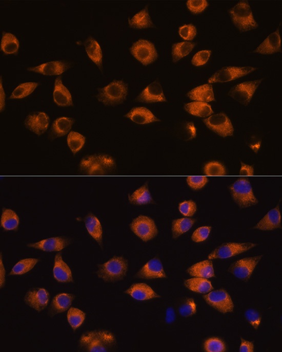
Immunofluorescence analysis of L929 cells using EPHA5 Rabbit pAb (CAB14238) at dilution of 1:100 (40x lens). Secondary antibody: Cy3-conjugated Goat anti-Rabbit IgG (H+L) (CABS007) at 1:500 dilution. Blue: DAPI for nuclear staining.