The ETFA Antibody (CAB7670) is a high-quality antibody developed for reliable detection and analysis of target proteins. This antibody, generated in rabbits, demonstrates strong reactivity with human samples and has been validated for use in Western blot applications.ETFA plays a crucial role in cellular energy metabolism and dysfunction in this protein has been linked to disorders like multiple acyl-CoA dehydrogenase deficiency. By targeting ETFA, researchers can gain insights into metabolic pathways and their implications in various diseases, making this antibody a valuable tool for studies in biochemistry, metabolism, and mitochondrial disorders.
This antibody is validated for use in WB, IHC-P, IP, ELISA applications and has demonstrated reactivity against Human, Mouse, Rat samples.
Product Name:
ETFA Antibody
SKU:
CAB7670
Size:
20μL, 100μL
Reactivity:
Human, Mouse, Rat
Conjugate:
Unconjugated
Immunogen:
Recombinant protein (or fragment).This information is considered to be commercially sensitive.
0.5μg-4μg antibody for 200μg-400μg extracts of whole cells
ELISA
Recommended starting concentration is 1 μg/mL. Please optimize the concentration based on your specific assay requirements.
Synonyms:
EMA, GA2, MADD, ETFA
Positive Sample:
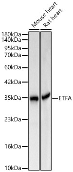
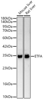
Mouse heart, Rat heart, Mouse liver, Rat liver
Cellular Localization:
Mitochondrion Matrix.
Calculated MW:
35kDa
Observed MW:
35kDa
ETFA participates in catalyzing the initial step of the mitochondrial fatty acid beta-oxidation. It shuttles electrons between primary flavoprotein dehydrogenases and the membrane-bound electron transfer flavoprotein ubiquinone oxidoreductase. Defects in electron-transfer-flavoprotein have been implicated in type II glutaricaciduria in which multiple acyl-CoA dehydrogenase deficiencies result in large excretion of glutaric, lactic, ethylmalonic, butyric, isobutyric, 2-methyl-butyric, and isovaleric acids. Two transcript variants encoding different isoforms have been found for this gene.
Purification Method
Affinity purification
Gene ID
2108
RRID
AB_2769366
Buffer Information
Store at -20℃. Avoid freeze / thaw cycles. Buffer: PBS containing 50% glycerol, preserved with proclin300 or sodium azide, pH 7.3.
Western blot analysis of various lysates using ETFA Rabbit pAb (CAB7670) at 1:1000 dilution. Secondary antibody: HRP-conjugated Goat anti-Rabbit IgG (H+L) (CABS014) at 1:10000 dilution. Lysates / proteins: 25 μg per lane. Blocking buffer: 3 % nonfat dry milk in TBST. Detection: ECL Basic Kit (AbGn00020). Exposure time: 1s.
Western blot analysis of various lysates using ETFA Rabbit pAb (CAB7670) at 1:1000 dilution. Secondary antibody: HRP-conjugated Goat anti-Rabbit IgG (H+L) (CABS014) at 1:10000 dilution. Lysates / proteins: 25 μg per lane. Blocking buffer: 3 % nonfat dry milk in TBST. Detection: ECL Basic Kit (AbGn00020). Exposure time: 10s.
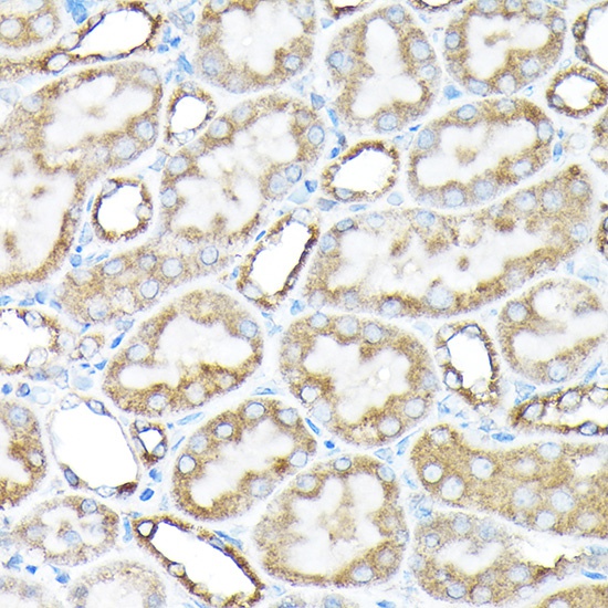
Immunohistochemistry analysis of paraffin-embedded Rat kidney using ETFA Rabbit pAb (CAB7670) at dilution of 1:200 (40x lens). High pressure antigen retrieval performed with 0.01M Citrate buffer (pH 6.0) prior to IHC staining.
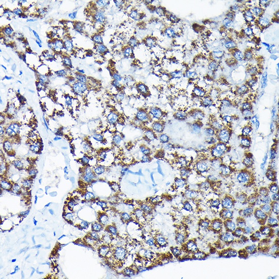
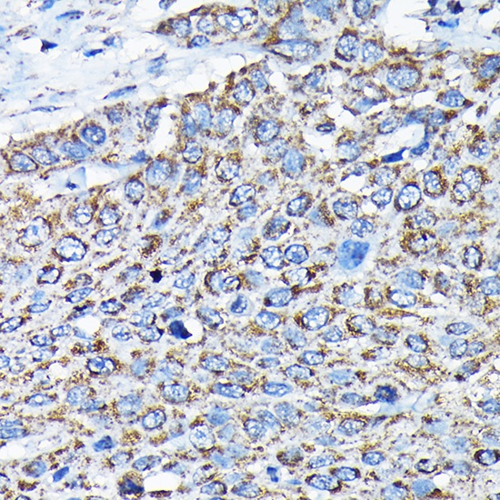
Immunohistochemistry analysis of paraffin-embedded Human liver cancer using ETFA Rabbit pAb (CAB7670) at dilution of 1:200 (40x lens). High pressure antigen retrieval performed with 0.01M Citrate buffer (pH 6.0) prior to IHC staining.
Immunohistochemistry analysis of paraffin-embedded Human esophageal cancer using ETFA Rabbit pAb (CAB7670) at dilution of 1:200 (40x lens). High pressure antigen retrieval performed with 0.01M Citrate buffer (pH 6.0) prior to IHC staining.
Immunohistochemistry analysis of paraffin-embedded Mouse spleen using ETFA Rabbit pAb (CAB7670) at dilution of 1:200 (40x lens). High pressure antigen retrieval performed with 0.01M Citrate buffer (pH 6.0) prior to IHC staining.
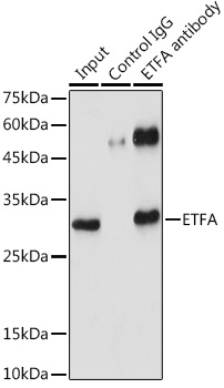
Immunoprecipitation analysis of 300 μg extracts of 293T cells using 3 μg ETFA antibody (CAB7670). Western blot was performed from the immunoprecipitate using ETFA antibody (CAB7670) at a dilution of 1:1000.