The ETHE1 Antibody (CAB10142) is a high-quality antibody developed for reliable detection and analysis of target proteins. This antibody, produced in rabbits, is highly specific for human samples and is validated for use in various applications, including Western blot and immunohistochemistry.The ETHE1 protein plays a crucial role in maintaining cellular homeostasis by catalyzing the conversion of persulfide to sulfide, a process essential for proper mitochondrial function and overall cellular health. Dysregulation of ETHE1 has been implicated in a variety of metabolic disorders and mitochondrial diseases, making it a promising target for therapeutic interventions.
This antibody is validated for use in WB, IHC-P, IF/ICC, ELISA applications and has demonstrated reactivity against Human, Mouse, Rat samples.
Product Name:
ETHE1 Antibody
SKU:
CAB10142
Size:
20μL, 100μL
Reactivity:
Human, Mouse, Rat
Conjugate:
Unconjugated
Immunogen:
Recombinant protein (or fragment).This information is considered to be commercially sensitive.
Recommended starting concentration is 1 μg/mL. Please optimize the concentration based on your specific assay requirements.
Synonyms:
HSCO, YF13H12, ETHE1
Positive Sample:
HT-29, HepG2, Mouse large intestine, Mouse liver, Rat liver
Cellular Localization:
Cytoplasm, Mitochondrion Matrix, Nucleus.
Calculated MW:
28kDa
Observed MW:
28kDa
This gene encodes a member of the metallo beta-lactamase family of iron-containing proteins involved in the mitochondrial sulfide oxidation pathway. The encoded protein catalyzes the oxidation of a persulfide substrate to sulfite. Certain mutations in this gene cause ethylmalonic encephalopathy, an infantile metabolic disorder affecting the brain, gastrointestinal tract and peripheral vessels. Alternative splicing results in multiple transcript variants encoding different isoforms.
Purification Method
Affinity purification
Gene ID
23474
RRID
AB_2757669
Buffer Information
Store at -20℃. Avoid freeze / thaw cycles. Buffer: PBS with 0.09% Sodium azide,50% glycerol,pH7.3.
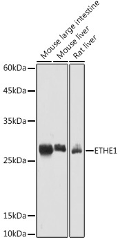
Western blot analysis of various lysates using ETHE1 Rabbit pAb (CAB10142) at 1:1000 dilution. Secondary antibody: HRP-conjugated Goat anti-Rabbit IgG (H+L) (CABS014) at 1:10000 dilution. Lysates/proteins: 25μg per lane. Blocking buffer: 3% nonfat dry milk in TBST. Detection: ECL Basic Kit (AbGn00020). Exposure time: 5s.
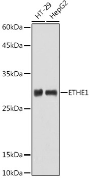
Western blot analysis of various lysates using ETHE1 Rabbit pAb (CAB10142) at 1:1000 dilution. Secondary antibody: HRP-conjugated Goat anti-Rabbit IgG (H+L) (CABS014) at 1:10000 dilution. Lysates/proteins: 25μg per lane. Blocking buffer: 3% nonfat dry milk in TBST. Detection: ECL Basic Kit (AbGn00020). Exposure time: 180s.
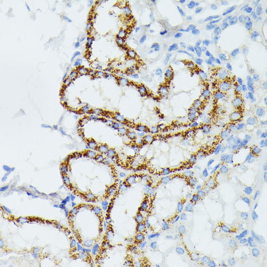
Immunohistochemistry analysis of paraffin-embedded Mouse kidney using ETHE1 Rabbit pAb (CAB10142) at dilution of 1:100 (40x lens). Microwave antigen retrieval performed with 0.01M PBS Buffer (pH 7.2) prior to IHC staining.
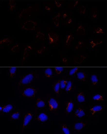
Immunofluorescence analysis of U-2 OS cells using ETHE1 Rabbit pAb (CAB10142) at dilution of 1:100 (40x lens). Secondary antibody: Cy3-conjugated Goat anti-Rabbit IgG (H+L) (CABS007) at 1:500 dilution. Blue: DAPI for nuclear staining.