The EWSR1 Monoclonal Antibody (CAB9640) is a high-quality antibody developed for reliable detection and analysis of target proteins. This antibody, derived from rabbits, exhibits high reactivity with human samples and is validated for use in immunohistochemistry and Western blot applications. By binding specifically to the EWSR1 protein, this antibody facilitates the detection and analysis of EWSR1 expression in different cell types, making it an ideal tool for studies in oncology and cancer biology.EWSR1, a key player in various oncogenic processes, is frequently involved in chromosomal translocations leading to the formation of oncogenic fusion proteins.
This antibody is validated for use in WB, IHC-P, IF/ICC, IP, ELISA applications and has demonstrated reactivity against Human, Mouse, Rat samples.
Product Name:
EWSR1 Monoclonal Antibody
SKU:
CAB9640
Size:
20μL, 100μL
Reactivity:
Human, Mouse, Rat
Clone Number:
ARC1674
Conjugate:
Unconjugated
Immunogen:
Recombinant protein (or fragment).This information is considered to be commercially sensitive.
0.5μg-4μg antibody for 200μg-400μg extracts of whole cells
ELISA
Recommended starting concentration is 1 μg/mL. Please optimize the concentration based on your specific assay requirements.
Synonyms:
EWS, EWS-FLI1, bK984G1.4, EWSR1
Positive Sample:
HeLa, Jurkat, Mouse testis, Mouse thymus, Rat testis
Cellular Localization:
Cell Membrane, Cytoplasm, Nucleus.
Calculated MW:
68kDa
Observed MW:
90kDa
This gene encodes a multifunctional protein that is involved in various cellular processes, including gene expression, cell signaling, and RNA processing and transport. The protein includes an N-terminal transcriptional activation domain and a C-terminal RNA-binding domain. Chromosomal translocations between this gene and various genes encoding transcription factors result in the production of chimeric proteins that are involved in tumorigenesis. These chimeric proteins usually consist of the N-terminal transcriptional activation domain of this protein fused to the C-terminal DNA-binding domain of the transcription factor protein. Mutations in this gene, specifically a t(11;22)(q24;q12) translocation, are known to cause Ewing sarcoma as well as neuroectodermal and various other tumors. Alternative splicing of this gene results in multiple transcript variants. Related pseudogenes have been identified on chromosomes 1 and 14.
Purification Method
Affinity purification
Gene ID
2130
RRID
AB_2863745
Buffer Information
Store at -20℃. Avoid freeze / thaw cycles. Buffer: PBS containing 50% glycerol and 0.05% BSA, preserved with proclin300 or sodium azide, pH 7.3.
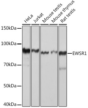
Western blot analysis of various lysates using EWSR1 Rabbit mAb (CAB9640) at 1:1000 dilution. Secondary antibody: HRP-conjugated Goat anti-Rabbit IgG (H+L) (CABS014) at 1:10000 dilution. Lysates/proteins: 25μg per lane. Blocking buffer: 3% nonfat dry milk in TBST. Detection: ECL Basic Kit (AbGn00020). Exposure time: 5s.
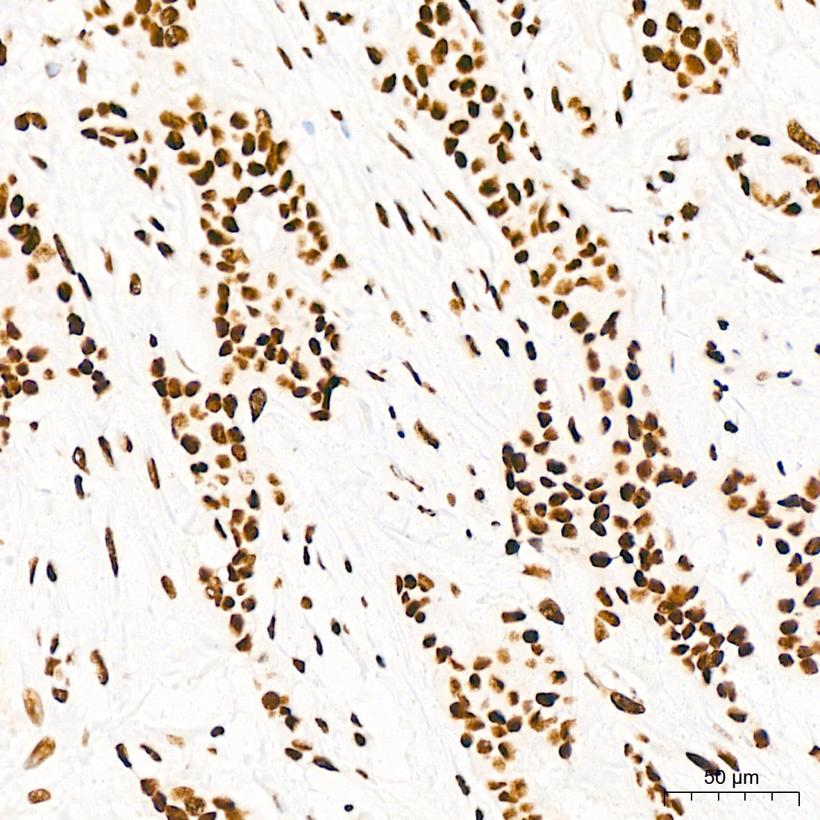
Immunohistochemistry analysis of paraffin-embedded Human breast cancer tissue using EWSR1 Rabbit mAb (CAB9640) at a dilution of 1:200 (40x lens). High pressure antigen retrieval performed with 0.01M Citrate buffer (pH 6.0) prior to IHC staining.
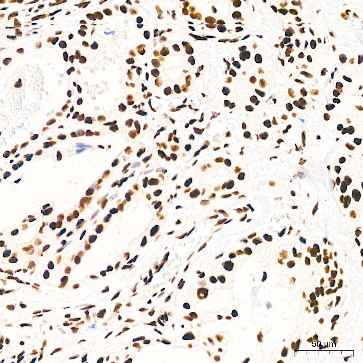
Immunohistochemistry analysis of paraffin-embedded Human thyroid tissue using EWSR1 Rabbit mAb (CAB9640) at a dilution of 1:200 (40x lens). High pressure antigen retrieval performed with 0.01M Citrate buffer (pH 6.0) prior to IHC staining.
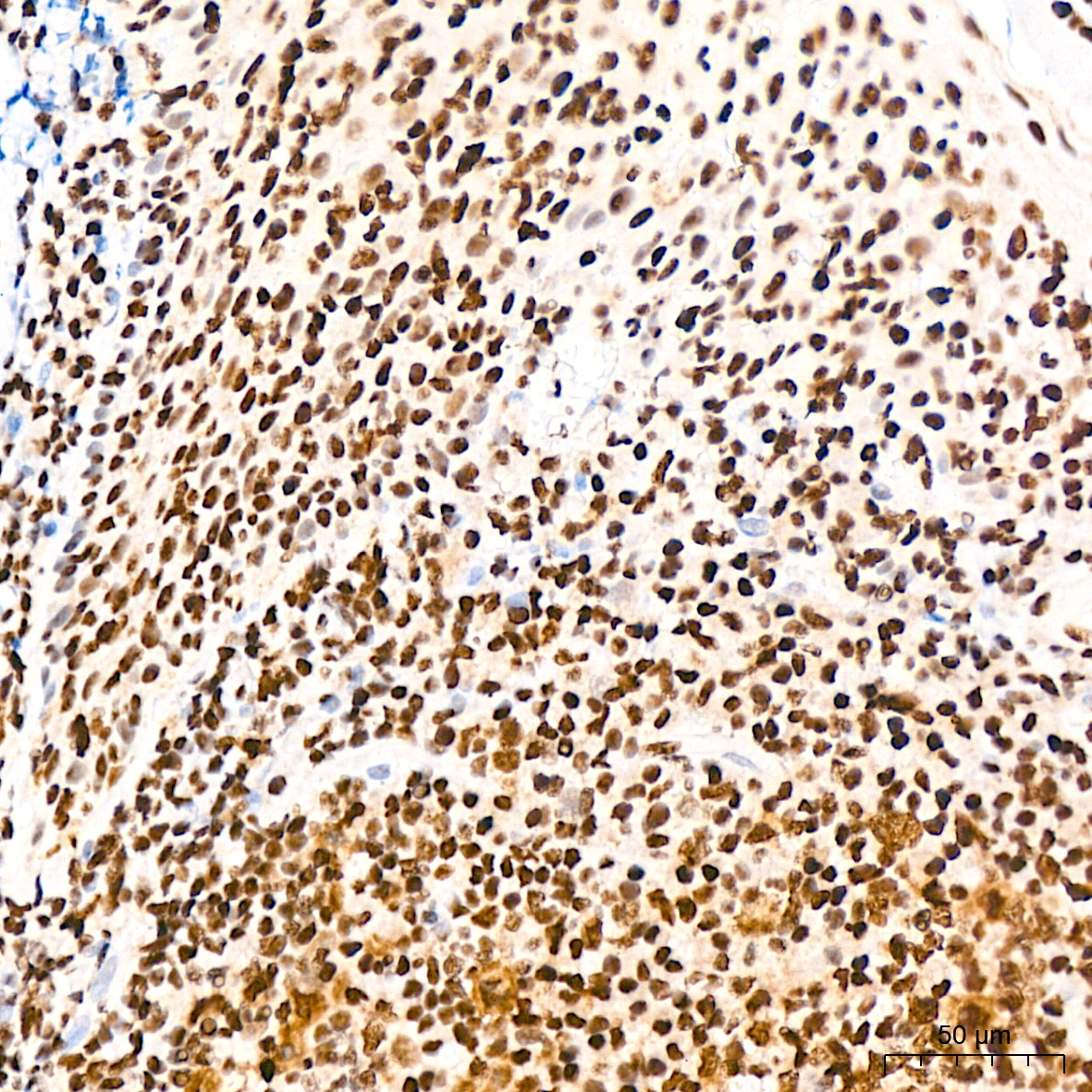
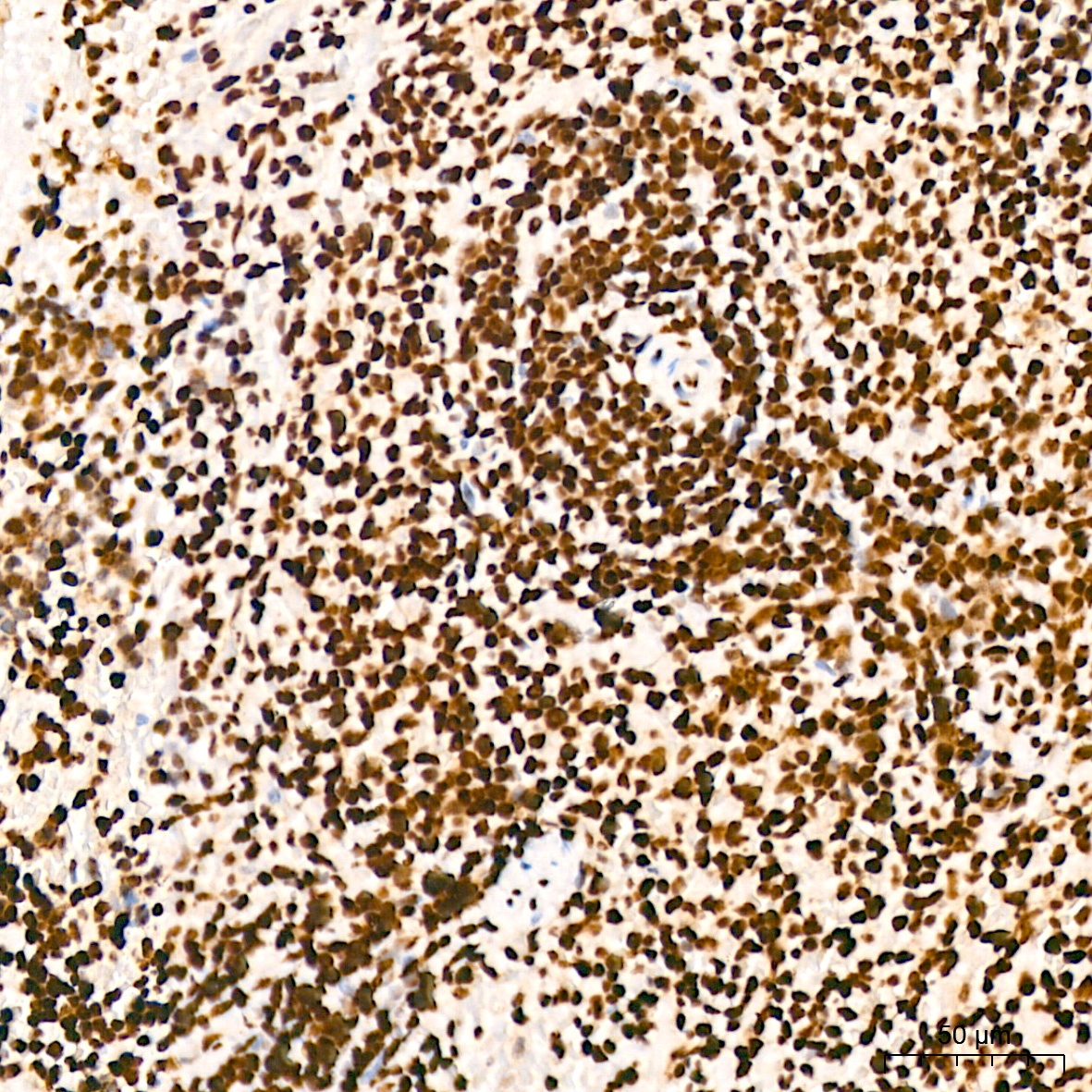
Immunohistochemistry analysis of paraffin-embedded Human tonsil tissue using EWSR1 Rabbit mAb (CAB9640) at a dilution of 1:200 (40x lens). High pressure antigen retrieval performed with 0.01M Citrate buffer (pH 6.0) prior to IHC staining.
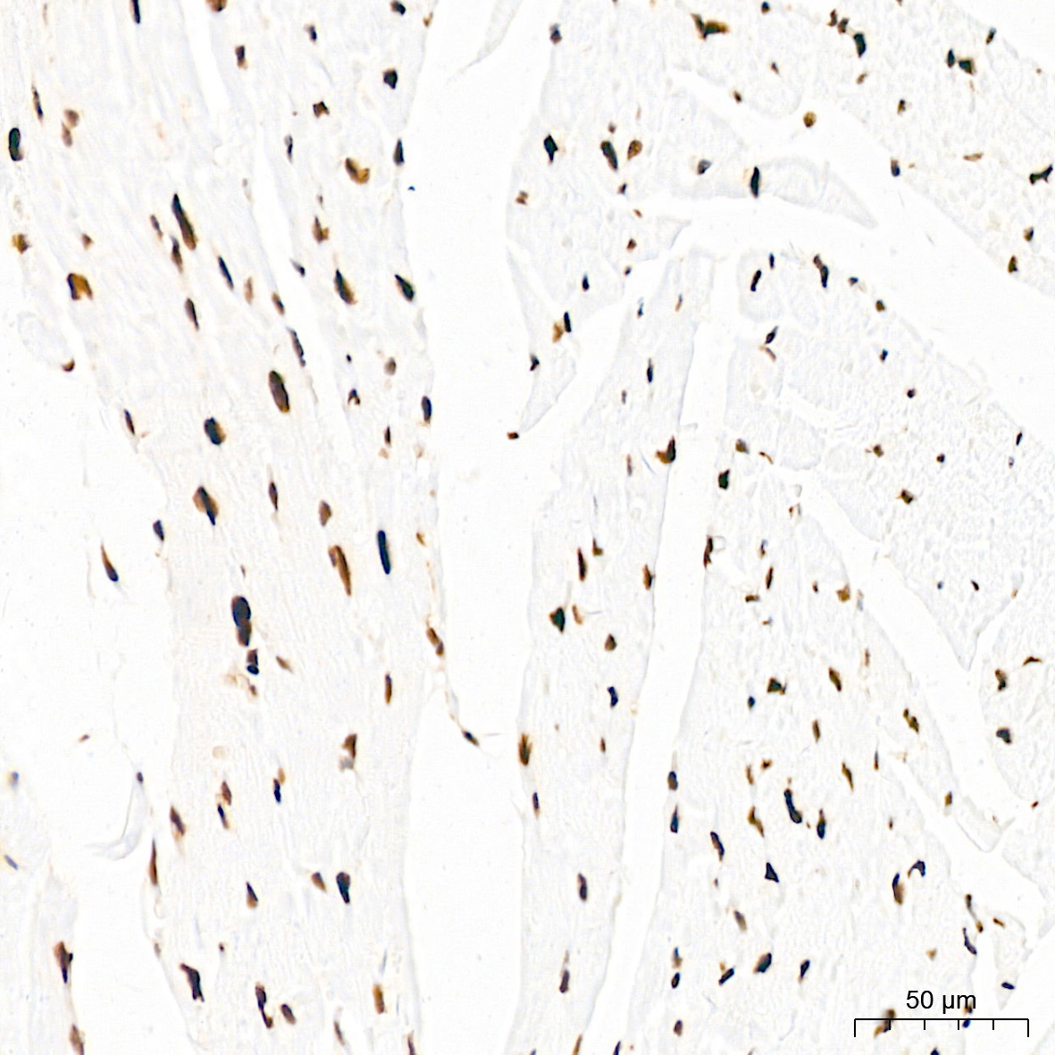
Immunohistochemistry analysis of paraffin-embedded Mouse heart tissue using EWSR1 Rabbit mAb (CAB9640) at a dilution of 1:200 (40x lens). High pressure antigen retrieval performed with 0.01M Citrate buffer (pH 6.0) prior to IHC staining.
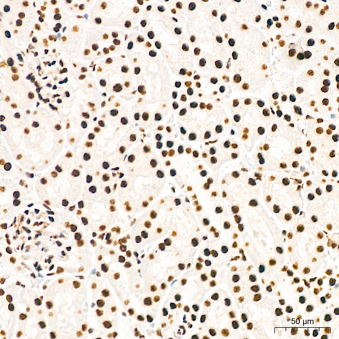
Immunohistochemistry analysis of paraffin-embedded Mouse kidney tissue using EWSR1 Rabbit mAb (CAB9640) at a dilution of 1:200 (40x lens). High pressure antigen retrieval performed with 0.01M Citrate buffer (pH 6.0) prior to IHC staining.
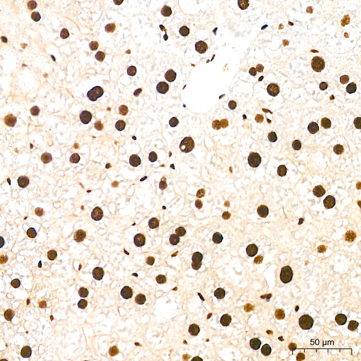
Immunohistochemistry analysis of paraffin-embedded Mouse liver tissue using EWSR1 Rabbit mAb (CAB9640) at a dilution of 1:200 (40x lens). High pressure antigen retrieval performed with 0.01M Citrate buffer (pH 6.0) prior to IHC staining.
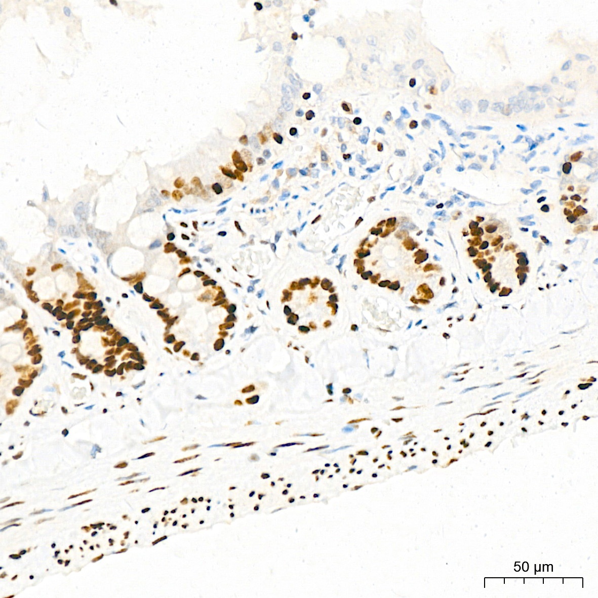
Immunohistochemistry analysis of paraffin-embedded Rat colon tissue using EWSR1 Rabbit mAb (CAB9640) at a dilution of 1:200 (40x lens). High pressure antigen retrieval performed with 0.01M Citrate buffer (pH 6.0) prior to IHC staining.
Immunohistochemistry analysis of paraffin-embedded Rat spleen tissue using EWSR1 Rabbit mAb (CAB9640) at a dilution of 1:200 (40x lens). High pressure antigen retrieval performed with 0.01M Citrate buffer (pH 6.0) prior to IHC staining.
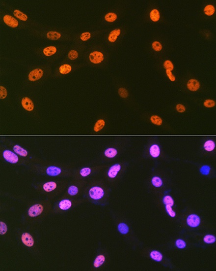
Immunofluorescence analysis of NIH-3T3 cells using EWSR1 Rabbit mAb (CAB9640) at dilution of 1:100 (40x lens). Secondary antibody: Cy3-conjugated Goat anti-Rabbit IgG (H+L) (CABS007) at 1:500 dilution. Blue: DAPI for nuclear staining.
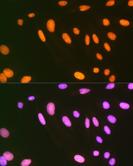
Immunofluorescence analysis of U-2 OS cells using EWSR1 Rabbit mAb (CAB9640) at dilution of 1:100 (40x lens). Secondary antibody: Cy3-conjugated Goat anti-Rabbit IgG (H+L) (CABS007) at 1:500 dilution. Blue: DAPI for nuclear staining.