The EXT2 Antibody (CAB1973) is a high-quality antibody developed for reliable detection and analysis of target proteins. This antibody, generated in rabbits, exhibits strong reactivity with human samples and has been validated for use in Western blot applications. By binding specifically to the Exostosin-2 protein, this antibody enables precise detection and analysis in a variety of cell types, making it an essential component for studies focusing on glycosaminoglycan biosynthesis and related pathways.Exostosin-2, also known as EXT2, plays a crucial role in heparan sulfate synthesis, which is essential for various biological processes such as cell signaling, development, and tissue organization.
This antibody is validated for use in WB, IF/ICC, ELISA applications and has demonstrated reactivity against Human, Mouse, Rat samples.
Product Name:
EXT2 Antibody
SKU:
CAB1973
Size:
20μL, 100μL
Reactivity:
Human, Mouse, Rat
Conjugate:
Unconjugated
Immunogen:
Recombinant protein (or fragment).This information is considered to be commercially sensitive.
Recommended starting concentration is 1 μg/mL. Please optimize the concentration based on your specific assay requirements.
Synonyms:
SOTV, SSMS, EXT2
Positive Sample:
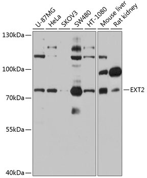
U-87MG, HeLa, SKOV3, SW480, HT-1080, Mouse liver, Rat kidney
Cellular Localization:
Endoplasmic Reticulum Membrane, Golgi Apparatus Membrane, Single-Pass Type Ii Membrane Protein.
Calculated MW:
82kDa
Observed MW:
82kDa
This gene encodes one of two glycosyltransferases involved in the chain elongation step of heparan sulfate biosynthesis. Mutations in this gene cause the type II form of multiple exostoses. Alternatively spliced transcript variants encoding different isoforms have been noted for this gene.
Purification Method
Affinity purification
Gene ID
2132
RRID
AB_2763999
Buffer Information
Store at -20℃. Avoid freeze / thaw cycles. Buffer: PBS containing 50% glycerol, preserved with proclin300 or sodium azide, pH 7.3.
Western blot analysis of various lysates using EXT2 Rabbit pAb (CAB1973) at 1:1000 dilution. Secondary antibody: HRP-conjugated Goat anti-Rabbit IgG (H+L) (CABS014) at 1:10000 dilution. Lysates/proteins: 25μg per lane. Blocking buffer: 3% nonfat dry milk in TBST. Detection: ECL Basic Kit (AbGn00020). Exposure time: 30s.
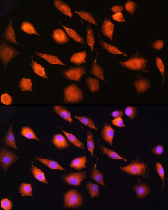
Immunofluorescence analysis of L929 cells using EXT2 Rabbit pAb (CAB1973) at dilution of 1:100 (40x lens). Secondary antibody: Cy3-conjugated Goat anti-Rabbit IgG (H+L) (CABS007) at 1:500 dilution. Blue: DAPI for nuclear staining.