The EYA3 Antibody (CAB15056) is a high-quality antibody developed for reliable detection and analysis of target proteins. This antibody, produced in rabbits, has been validated for use in Western blot applications and demonstrates high reactivity with human samples.EYA3 is a member of the EYA family of proteins, known for their role in mediating protein-protein interactions and acting as transcriptional co-activators.
This antibody is validated for use in WB, IF/ICC, ELISA applications and has demonstrated reactivity against Human, Rat samples.
Product Name:
EYA3 Antibody
SKU:
CAB15056
Size:
20μL, 100μL
Reactivity:
Human, Rat
Conjugate:
Unconjugated
Immunogen:
Recombinant protein (or fragment).This information is considered to be commercially sensitive.
Recommended starting concentration is 1 μg/mL. Please optimize the concentration based on your specific assay requirements.
Synonyms:
EYA3
Positive Sample:
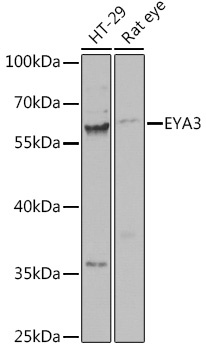
HT-29, Rat eye
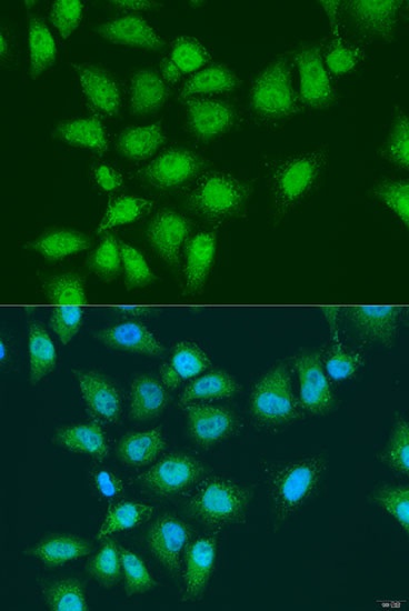
Cellular Localization:
Cytoplasm, Nucleus.
Calculated MW:
63kDa
Observed MW:
57kDa/58kDa
This gene encodes a member of the eyes absent (EYA) family of proteins. The encoded protein may act as a transcriptional activator and have a role during development. It can act as a mediator of chemoresistance and cell survival in Ewing sarcoma cells, where this gene is up-regulated via a micro-RNA that binds to the 3' UTR of the transcript. A similar protein in mice acts as a transcriptional activator. Alternative splicing of this gene results in multiple transcript variants.
Purification Method
Affinity purification
Gene ID
2140
RRID
AB_2761937
Buffer Information
Store at -20℃. Avoid freeze / thaw cycles. Buffer: PBS containing 50% glycerol, preserved with proclin300 or sodium azide, pH 7.3.
Western blot analysis of various lysates using EYA3 Rabbit pAb (CAB15056) at 1:1000 dilution. Secondary antibody: HRP-conjugated Goat anti-Rabbit IgG (H+L) (CABS014) at 1:10000 dilution. Lysates/proteins: 25μg per lane. Blocking buffer: 3% nonfat dry milk in TBST. Detection: ECL Basic Kit (AbGn00020). Exposure time: 30s.
Immunofluorescence analysis of U2OS cells using EYA3 Rabbit pAb (CAB15056) at dilution of 1:100. Secondary antibody: Cy3-conjugated Goat anti-Rabbit IgG (H+L) (CABS007) at 1:500 dilution. Blue: DAPI for nuclear staining.