The FABP1 Antibody (CAB5311) is a high-quality antibody developed for reliable detection and analysis of target proteins. This antibody is raised in rabbits and has high reactivity with human samples, making it ideal for various research applications. Validated for use in Western blot analyses, this antibody binds to the FABP1 protein, allowing for accurate detection and analysis in different cell types.FABP1 is a crucial protein involved in lipid metabolism and transport within the liver.
This antibody is validated for use in WB, IF/ICC, ELISA applications and has demonstrated reactivity against Human, Mouse, Rat samples.
Product Name:
FABP1 Antibody
SKU:
CAB5311
Size:
20μL, 100μL
Reactivity:
Human, Mouse, Rat
Conjugate:
Unconjugated
Immunogen:
Recombinant protein (or fragment).This information is considered to be commercially sensitive.
Recommended starting concentration is 1 μg/mL. Please optimize the concentration based on your specific assay requirements.
Synonyms:
FABPL, L-FABP, FABP1
Positive Sample:
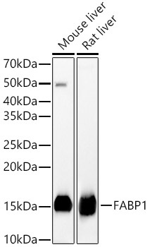
Mouse liver, Rat liver
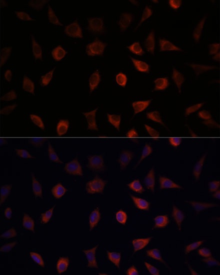
Cellular Localization:
Cytoplasm.
Calculated MW:
14kDa
Observed MW:
14kDa
This gene encodes the fatty acid binding protein found in liver. Fatty acid binding proteins are a family of small, highly conserved, cytoplasmic proteins that bind long-chain fatty acids and other hydrophobic ligands. This protein and FABP6 (the ileal fatty acid binding protein) are also able to bind bile acids. It is thought that FABPs roles include fatty acid uptake, transport, and metabolism.
Purification Method
Affinity purification
Gene ID
2168
RRID
AB_2766123
Buffer Information
Store at -20℃. Avoid freeze / thaw cycles. Buffer: PBS containing 50% glycerol, preserved with proclin300 or sodium azide, pH 7.3.
Western blot analysis of various lysates, using FABP1 Rabbit pAb (CAB5311) at 1:5000 dilution. Secondary antibody: HRP-conjugated Goat anti-Rabbit IgG (H+L) (CABS014) at 1:10000 dilution. Lysates/proteins: 25μg per lane. Blocking buffer: 3% nonfat dry milk in TBST. Detection: ECL Basic Kit (AbGn00020). Exposure time: 30s.
Immunofluorescence analysis of L929 cells using FABP1 Rabbit pAb (CAB5311) at dilution of 1:100. Secondary antibody: Cy3-conjugated Goat anti-Rabbit IgG (H+L) (CABS007) at 1:500 dilution. Blue: DAPI for nuclear staining.