The FAM107A Antibody (CAB16494) is a high-quality antibody developed for reliable detection and analysis of target proteins. This antibody, produced in rabbits, exhibits high reactivity with human samples and has been validated for use in Western blot applications. By binding specifically to the FAM107A protein, this antibody enables accurate detection and analysis in a variety of cell types, making it an ideal choice for studies in molecular biology and cell signaling.FAM107A, also known as family with sequence similarity 107 member A, is implicated in diverse biological functions, including regulation of cell growth, differentiation, and apoptosis.
This antibody is validated for use in WB, ELISA applications and has demonstrated reactivity against Human, Mouse samples.
Product Name:
FAM107A Antibody
SKU:
CAB16494
Size:
20μL, 100μL
Reactivity:
Human, Mouse
Conjugate:
Unconjugated
Immunogen:
Recombinant protein (or fragment).This information is considered to be commercially sensitive.
Recommended starting concentration is 1 μg/mL. Please optimize the concentration based on your specific assay requirements.
Synonyms:
DRR1, TU3A, FAM107A
Positive Sample:
K-562
Cellular Localization:
Nucleus.
Calculated MW:
17kDa
Observed MW:
18kDa
Predicted to enable actin binding activity. Involved in several processes, including negative regulation of G1/S transition of mitotic cell cycle; negative regulation of focal adhesion assembly; and regulation of cytoskeleton organization. Located in several cellular components, including focal adhesion; ruffle membrane; and stress fiber.
Purification Method
Affinity purification
Gene ID
11170
RRID
AB_2769402
Buffer Information
Store at -20℃. Avoid freeze / thaw cycles. Buffer: PBS with 0.01% thimerosal,50% glycerol,pH7.3.
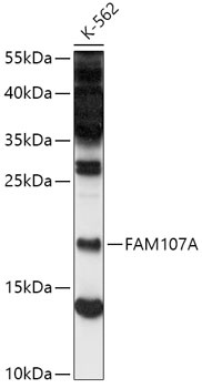
Western blot analysis of lysates from K-562 cells, using FAM107A Rabbit pAb (CAB16494) at 1:500 dilution. Secondary antibody: HRP-conjugated Goat anti-Rabbit IgG (H+L) (CABS014) at 1:10000 dilution. Lysates/proteins: 25μg per lane. Blocking buffer: 3% nonfat dry milk in TBST. Detection: ECL Basic Kit (AbGn00020). Exposure time: 60s.