The FBXW11 Antibody (CAB7784) is a high-quality antibody developed for reliable detection and analysis of target proteins. This antibody, produced in rabbits, exhibits high specificity and sensitivity for human samples, making it an ideal choice for Western blot applications.FBXW11, also known as F-box/WD repeat-containing protein 11, is a crucial component of the ubiquitin-proteasome system, a pathway that controls the turnover of proteins involved in cell growth and division. Dysregulation of FBXW11 has been implicated in various diseases, including cancer and neurological disorders, highlighting its importance as a potential therapeutic target.
This antibody is validated for use in WB, IF/ICC, ELISA applications and has demonstrated reactivity against Human, Mouse, Rat samples.
Product Name:
FBXW11 Antibody
SKU:
CAB7784
Size:
20μL, 100μL
Reactivity:
Human, Mouse, Rat
Conjugate:
Unconjugated
Immunogen:
Synthetic peptide. This information is considered to be commercially sensitive.
Sequence:
FLNV PPSA QNET RS
Tested Applications:
WBIF/ICCELISA
Recommended Dilution:
WB
1:500 - 1:1000
IF/ICC
1:50 - 1:200
ELISA
Recommended starting concentration is 1 μg/mL. Please optimize the concentration based on your specific assay requirements.
This gene encodes a member of the F-box protein family which is characterized by an approximately 40 amino acid motif, the F-box. The F-box proteins constitute one of the four subunits of ubiquitin protein ligase complex called SCFs (SKP1-cullin-F-box), which function in phosphorylation-dependent ubiquitination. The F-box proteins are divided into 3 classes: Fbws containing WD-40 domains, Fbls containing leucine-rich repeats, and Fbxs containing either different protein-protein interaction modules or no recognizable motifs. The protein encoded by this gene belongs to the Fbws class and, in addition to an F-box, contains multiple WD40 repeats. This gene contains at least 14 exons, and its alternative splicing generates 3 transcript variants diverging at the presence/absence of two alternate exons.
Purification Method
Affinity purification
Gene ID
23291
RRID
AB_2769436
Buffer Information
Store at -20℃. Avoid freeze / thaw cycles. Buffer: PBS containing 50% glycerol, preserved with proclin300 or sodium azide, pH 7.3.
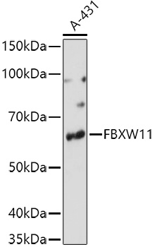
Western blot analysis of lysates from A-431 cells, using FBXW11 Rabbit pAb (CAB7784) at 1:1000 dilution. Secondary antibody: HRP-conjugated Goat anti-Rabbit IgG (H+L) (CABS014) at 1:10000 dilution. Lysates/proteins: 25μg per lane. Blocking buffer: 3% nonfat dry milk in TBST. Detection: ECL Basic Kit (AbGn00020). Exposure time: 90s.
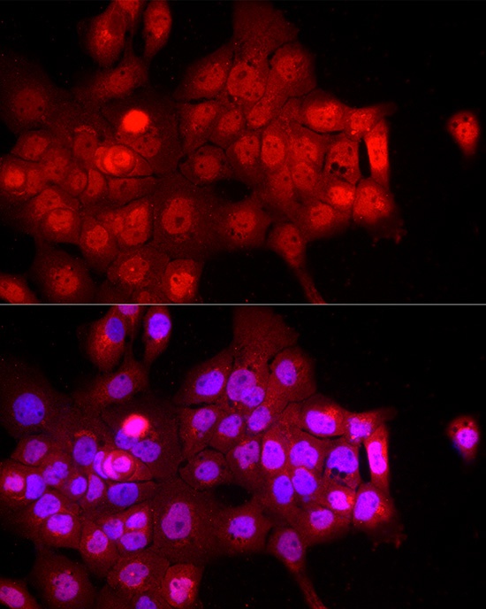
Immunofluorescence analysis of A-431 cells using FBXW11 Rabbit pAb (CAB7784) at dilution of 1:200 (40x lens). Secondary antibody: Cy3-conjugated Goat anti-Rabbit IgG (H+L) (CABS007) at 1:500 dilution. Blue: DAPI for nuclear staining.
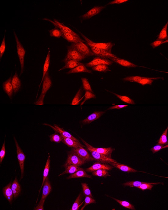
Immunofluorescence analysis of NIH/3T3 cells using FBXW11 Rabbit pAb (CAB7784) at dilution of 1:200 (40x lens). Secondary antibody: Cy3-conjugated Goat anti-Rabbit IgG (H+L) (CABS007) at 1:500 dilution. Blue: DAPI for nuclear staining.
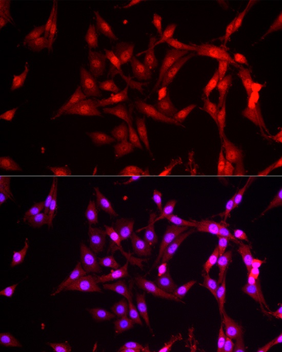
Immunofluorescence analysis of PC-12 cells using FBXW11 Rabbit pAb (CAB7784) at dilution of 1:200 (40x lens). Secondary antibody: Cy3-conjugated Goat anti-Rabbit IgG (H+L) (CABS007) at 1:500 dilution. Blue: DAPI for nuclear staining.