The FCF1 Antibody (CAB17149) is a high-quality antibody developed for reliable detection and analysis of target proteins. This antibody, generated in rabbits, is highly specific and reactive with human samples, making it suitable for various applications such as Western blot and immunohistochemistry.FCF1 is known to play a crucial role in gene expression, particularly in the regulation of mRNA splicing and processing. Its involvement in these processes makes it a key player in the control of gene expression and cellular functions. Researchers studying RNA biology, transcription regulation, or gene expression will find this antibody particularly useful in their experiments.
This antibody is validated for use in WB, IHC-P, IF/ICC, ELISA applications and has demonstrated reactivity against Human, Rat samples.
Product Name:
FCF1 Antibody
SKU:
CAB17149
Size:
20μL, 100μL
Reactivity:
Human, Rat
Immunogen:
Synthetic peptide. This information is considered to be commercially sensitive.
Recommended starting concentration is 1 μg/mL. Please optimize the concentration based on your specific assay requirements.
Synonyms:
Bka, UTP24, CGI-35, C14orf111, FCF1
Positive Sample:
293T
Cellular Localization:
Nucleolus, Nucleoplasm.
Calculated MW:
23kDa
Observed MW:
23kDa
Enables RNA binding activity. Predicted to be involved in rRNA processing. Predicted to act upstream of or within endonucleolytic cleavage in 5'-ETS of tricistronic rRNA transcript (SSU-rRNA, 5.8S rRNA, LSU-rRNA) and endonucleolytic cleavage in ITS1 to separate SSU-rRNA from 5.8S rRNA and LSU-rRNA from tricistronic rRNA transcript (SSU-rRNA, 5.8S rRNA, LSU-rRNA). Predicted to be located in nucleoplasm. Predicted to be part of small-subunit processome. Predicted to be active in nucleolus.
Purification Method
Affinity purification
Gene ID
51077
RRID
AB_2769441
Buffer Information
Store at -20℃. Avoid freeze / thaw cycles. Buffer: PBS with 0.01% thimerosal,50% glycerol,pH7.3.
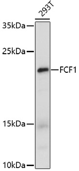
Western blot analysis of lysates from 293T cells, using FCF1 Rabbit pAb (CAB17149) at 1:1000 dilution. Secondary antibody: HRP-conjugated Goat anti-Rabbit IgG (H+L) (CABS014) at 1:10000 dilution. Lysates/proteins: 25μg per lane. Blocking buffer: 3% nonfat dry milk in TBST. Detection: ECL Enhanced Kit (AbGn00021). Exposure time: 100s.
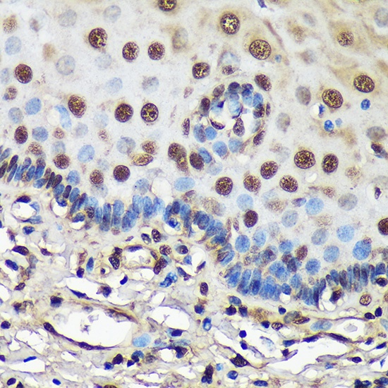
Immunohistochemistry analysis of paraffin-embedded Human esophageal using FCF1 Rabbit pAb (CAB17149) at dilution of 1:100 (40x lens). Microwave antigen retrieval performed with 0.01M PBS Buffer (pH 7.2) prior to IHC staining.
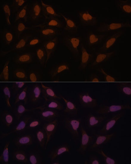
Immunofluorescence analysis of U-2 OS cells using FCF1 Rabbit pAb (CAB17149) at dilution of 1:100. Secondary antibody: Cy3-conjugated Goat anti-Rabbit IgG (H+L) (CABS007) at 1:500 dilution. Blue: DAPI for nuclear staining.