The FGFR1OP Antibody (CAB17627) is a high-quality antibody developed for reliable detection and analysis of target proteins. Raised in rabbits, this antibody is highly specific for human samples and is suitable for use in various applications, including Western blot and immunohistochemistry.FGFR1 is a crucial component in cellular signaling pathways involved in development, tissue repair, and disease progression. Dysregulation of FGFR1 has been implicated in a variety of cancers, making it an attractive target for cancer research.
This antibody is validated for use in WB, ELISA applications and has demonstrated reactivity against Mouse, Rat samples.
Product Name:
FGFR1OP Antibody
SKU:
CAB17627
Size:
20μL, 100μL
Reactivity:
Mouse, Rat
Conjugate:
Unconjugated
Immunogen:
Recombinant protein (or fragment).This information is considered to be commercially sensitive.
This gene encodes a largely hydrophilic centrosomal protein that is required for anchoring microtubules to subcellular structures. A t(6;8)(q27;p11) chromosomal translocation, fusing this gene and the fibroblast growth factor receptor 1 (FGFR1) gene, has been found in cases of myeloproliferative disorder. The resulting chimeric protein contains the N-terminal leucine-rich region of this encoded protein fused to the catalytic domain of FGFR1. Alterations in this gene may also be associated with Crohn's disease, Graves' disease, and vitiligo. Alternatively spliced transcript variants that encode different proteins have been identified.
Purification Method
Affinity purification
Gene ID
11116
RRID
AB_2769468
Buffer Information
Store at -20℃. Avoid freeze / thaw cycles. Buffer: PBS with 0.01% thimerosal,50% glycerol,pH7.3.
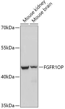
Western blot analysis of various lysates using FGFR1OP Rabbit pAb (CAB17627) at 1:1000 dilution. Secondary antibody: HRP-conjugated Goat anti-Rabbit IgG (H+L) (CABS014) at 1:10000 dilution. Lysates/proteins: 25μg per lane. Blocking buffer: 3% nonfat dry milk in TBST. Detection: ECL Basic Kit (AbGn00020). Exposure time: 90s.