The FGR Antibody (CAB2075) is a high-quality antibody developed for reliable detection and analysis of target proteins. This antibody, produced in rabbits, exhibits high specificity and sensitivity for detecting FGR in human samples, making it an essential component in Western blot applications. By specifically targeting the FGR protein, this antibody enables precise detection and analysis in a variety of cell types, making it suitable for investigations in immunology, cancer research, and beyond.FGR is a key player in cellular signaling pathways, particularly those involved in regulating cell growth, differentiation, and survival. Its role in oncogenesis and immune responses has garnered significant interest in the scientific community, as dysregulation of FGR has been implicated in various disease states, including cancer and inflammatory disorders.
This antibody is validated for use in WB, IHC-P, ELISA applications and has demonstrated reactivity against Human, Rat samples.
Product Name:
FGR Antibody
SKU:
CAB2075
Size:
20μL, 100μL
Reactivity:
Human, Rat
Conjugate:
Unconjugated
Immunogen:
Synthetic peptide. This information is considered to be commercially sensitive.
This gene is a member of the Src family of protein tyrosine kinases (PTKs). The encoded protein contains N-terminal sites for myristylation and palmitylation, a PTK domain, and SH2 and SH3 domains which are involved in mediating protein-protein interactions with phosphotyrosine-containing and proline-rich motifs, respectively. The protein localizes to plasma membrane ruffles, and functions as a negative regulator of cell migration and adhesion triggered by the beta-2 integrin signal transduction pathway. Infection with Epstein-Barr virus results in the overexpression of this gene. Multiple alternatively spliced variants, encoding the same protein, have been identified.
Purification Method
Affinity purification
Gene ID
2268
RRID
AB_2764097
Buffer Information
Store at -20℃. Avoid freeze / thaw cycles. Buffer: PBS containing 50% glycerol, preserved with proclin300 or sodium azide, pH 7.3.
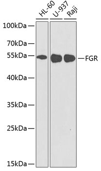
Western blot analysis of various lysates using FGR Rabbit pAb (CAB2075) at 1:1000 dilution._Secondary antibody: HRP-conjugated Goat anti-Rabbit IgG (H+L) (CABS014) at 1:10000 dilution._Lysates/proteins: 25μg per lane._Blocking buffer: 3% nonfat dry milk in TBST._Detection: ECL Enhanced Kit (AbGn00021)._Exposure time: 90s.
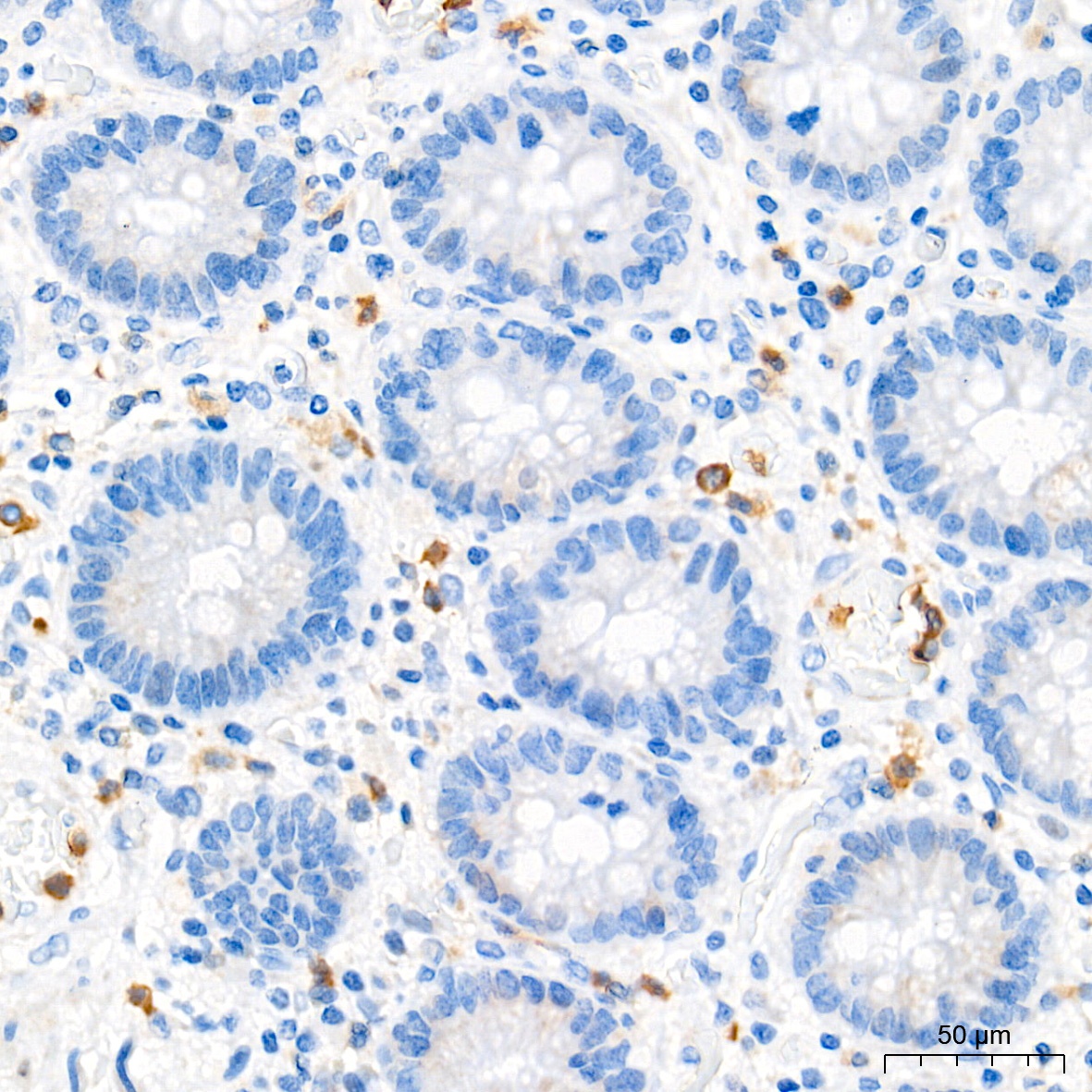
Immunohistochemistry analysis of paraffin-embedded Human colon tissue using FGR Rabbit pAb (CAB2075) at a dilution of 1:100 (40x lens). High pressure antigen retrieval performed with 0.01M Tris-EDTA Buffer (pH 9.0) prior to IHC staining.
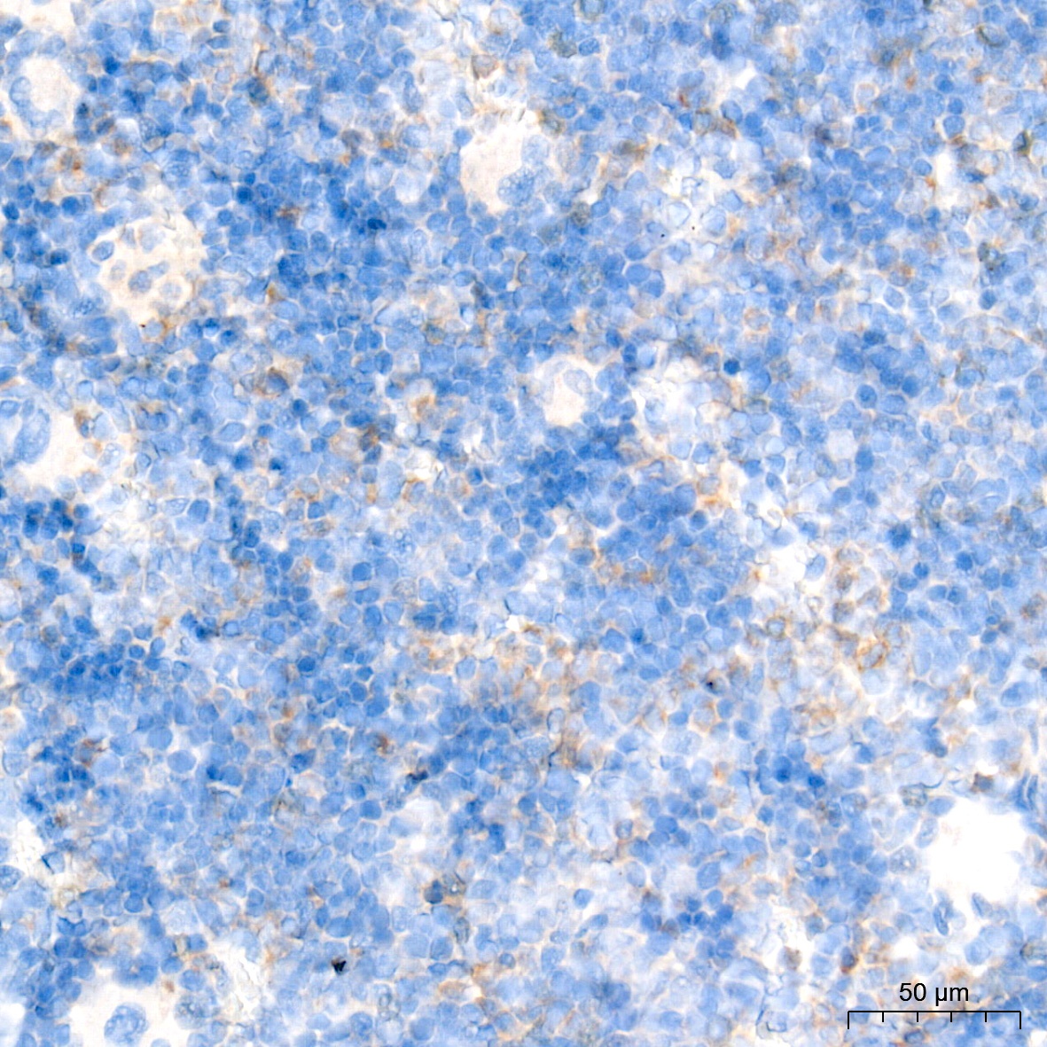
Immunohistochemistry analysis of paraffin-embedded Rat bone marrow tissue using FGR Rabbit pAb (CAB2075) at a dilution of 1:100 (40x lens). High pressure antigen retrieval performed with 0.01M Tris-EDTA Buffer (pH 9.0) prior to IHC staining.
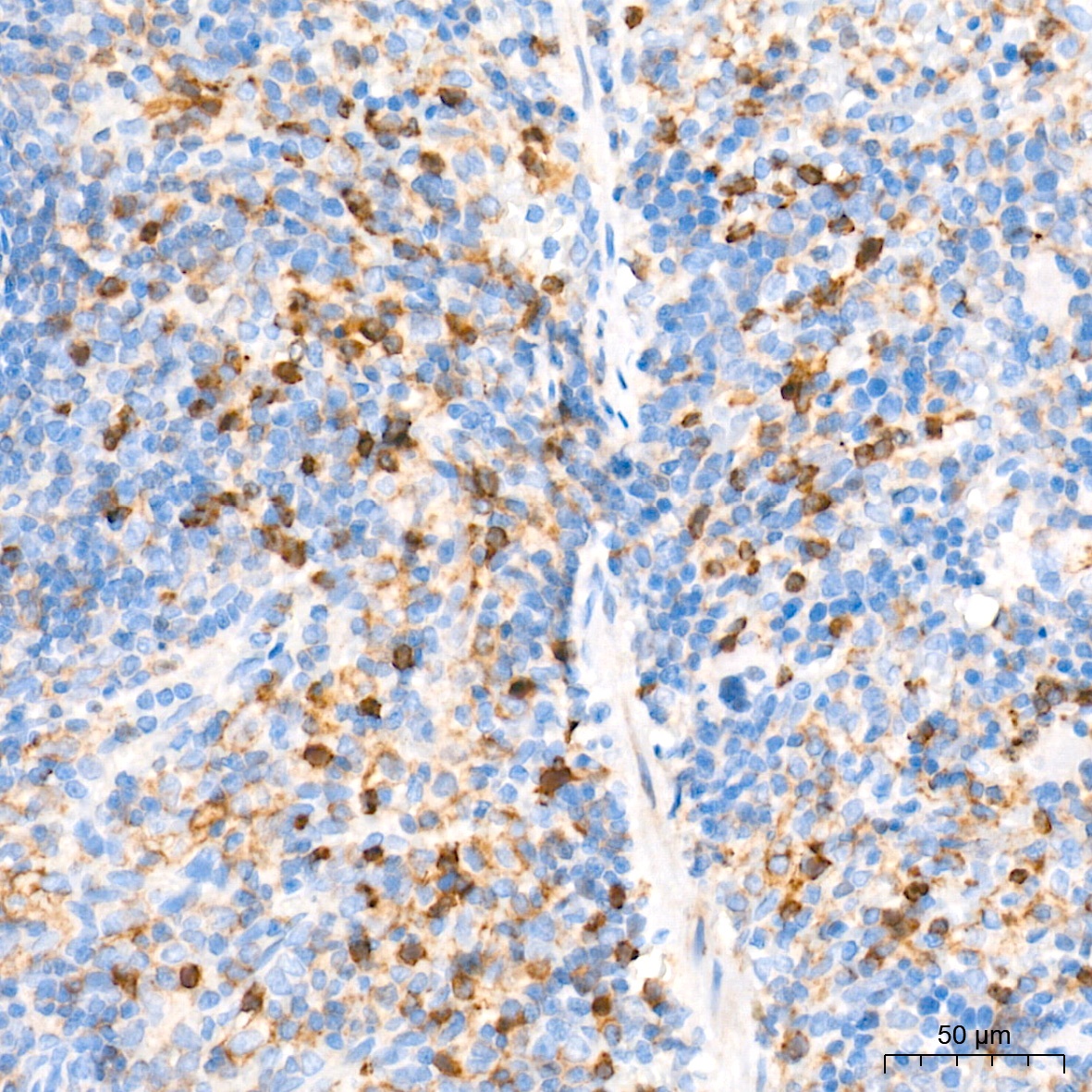
Immunohistochemistry analysis of paraffin-embedded Rat spleen tissue using FGR Rabbit pAb (CAB2075) at a dilution of 1:100 (40x lens). High pressure antigen retrieval performed with 0.01M Tris-EDTA Buffer (pH 9.0) prior to IHC staining.