The FH Antibody (CAB5688) is a high-quality antibody developed for reliable detection and analysis of target proteins. The antibody, raised in rabbits, exhibits high reactivity with human samples and has been validated for use in Western blot applications. By binding to the FH protein, this antibody enables efficient detection and analysis in various cell types, making it an invaluable tool for studies in immunology and complement-mediated diseases.The FH protein plays a crucial role in preventing excessive activation of the complement system and protecting host cells from damage. Dysregulation of FH function has been implicated in various diseases, including autoimmune disorders, inflammatory conditions, and certain kidney diseases.
This antibody is validated for use in WB, IHC-P, IF/ICC, ELISA applications and has demonstrated reactivity against Human, Mouse, Rat samples.
Product Name:
FH Antibody
SKU:
CAB5688
Size:
20μL, 100μL
Reactivity:
Human, Mouse, Rat
Conjugate:
Unconjugated
Immunogen:
Recombinant protein (or fragment).This information is considered to be commercially sensitive.
Recommended starting concentration is 1 μg/mL. Please optimize the concentration based on your specific assay requirements.
Synonyms:
MCL, FMRD, HsFH, LRCC, HLRCC, MCUL1, FH
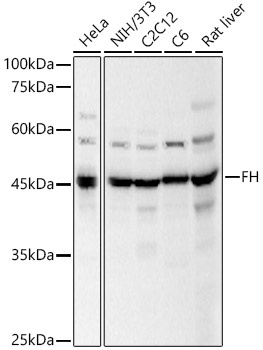
Positive Sample:
HeLa, NIH/3T3, C2C12, C6, Rat liver
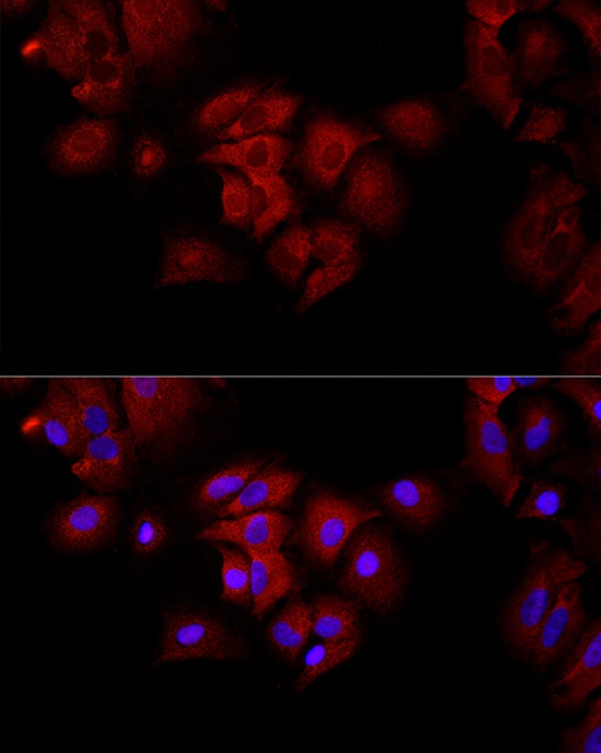
Cellular Localization:
Cytoplasm, Mitochondrion.
Calculated MW:
55kDa
Observed MW:
47kDa
The protein encoded by this gene is an enzymatic component of the tricarboxylic acid (TCA) cycle, or Krebs cycle, and catalyzes the formation of L-malate from fumarate. It exists in both a cytosolic form and an N-terminal extended form, differing only in the translation start site used. The N-terminal extended form is targeted to the mitochondrion, where the removal of the extension generates the same form as in the cytoplasm. It is similar to some thermostable class II fumarases and functions as a homotetramer. Mutations in this gene can cause fumarase deficiency and lead to progressive encephalopathy.
Purification Method
Affinity purification
Gene ID
2271
RRID
AB_2766448
Buffer Information
Store at -20℃. Avoid freeze / thaw cycles. Buffer: PBS containing 50% glycerol, preserved with proclin300 or sodium azide, pH 7.3.
Western blot analysis of various lysates, using FH Rabbit pAb (CAB5688) at 1:500 dilution. Secondary antibody: HRP-conjugated Goat anti-Rabbit IgG (H+L) (CABS014) at 1:10000 dilution. Lysates/proteins: 25μg per lane. Blocking buffer: 3% nonfat dry milk in TBST. Detection: ECL Basic Kit (AbGn00020). Exposure time: 5s.
Immunofluorescence analysis of A-549 cells using FH Rabbit pAb (CAB5688) at dilution of 1:50 (40x lens). Secondary antibody: Cy3-conjugated Goat anti-Rabbit IgG (H+L) (CABS007) at 1:500 dilution. Blue: DAPI for nuclear staining.