The FIP1L1 Antibody (CAB7138) is a high-quality antibody developed for reliable detection and analysis of target proteins. This antibody, generated in rabbits, exhibits high specificity and sensitivity when used with human samples, making it an excellent choice for Western blot applications. By targeting the FIP1L1 protein, researchers can accurately detect and study its function in different cell types, making it ideal for investigations in molecular biology and genetics.FIP1L1 is a key component in the formation of the pre-mRNA 3' processing complex, playing a critical role in gene transcription and RNA processing.
This antibody is validated for use in WB, IHC-P, IF/ICC, ELISA applications and has demonstrated reactivity against Human, Mouse, Rat samples.
Product Name:
FIP1L1 Antibody
SKU:
CAB7138
Size:
20μL, 100μL
Reactivity:
Human, Mouse, Rat
Conjugate:
Unconjugated
Immunogen:
Recombinant protein (or fragment).This information is considered to be commercially sensitive.
Recommended starting concentration is 1 μg/mL. Please optimize the concentration based on your specific assay requirements.
Synonyms:
Rhe, FIP1, hFip1, FIP1L1
Positive Sample:
Jurkat, 293F, Mouse thymus, Mouse brain
Cellular Localization:
Nucleus.
Calculated MW:
67kDa
Observed MW:
75kDa
This gene encodes a subunit of the CPSF (cleavage and polyadenylation specificity factor) complex that polyadenylates the 3' end of mRNA precursors. This gene, the homolog of yeast Fip1 (factor interacting with PAP), binds to U-rich sequences of pre-mRNA and stimulates poly(A) polymerase activity. Its N-terminus contains a PAP-binding site and its C-terminus an RNA-binding domain. An interstitial chromosomal deletion on 4q12 creates an in-frame fusion of human genes FIP1L1 and PDGFRA (platelet-derived growth factor receptor, alpha). The FIP1L1-PDGFRA fusion gene encodes a constitutively activated tyrosine kinase that joins the first 233 amino acids of FIP1L1 to the last 523 amino acids of PDGFRA. This gene fusion and chromosomal deletion is the cause of some forms of idiopathic hypereosinophilic syndrome (HES). This syndrome, recently reclassified as chronic eosinophilic leukemia (CEL), is responsive to treatment with tyrosine kinase inhibitors. Alternative splicing results in multiple transcript variants encoding distinct isoforms.
Purification Method
Affinity purification
Gene ID
81608
RRID
AB_2767693
Buffer Information
Store at -20℃. Avoid freeze / thaw cycles. Buffer: PBS containing 50% glycerol, preserved with proclin300 or sodium azide, pH 7.3.
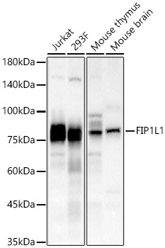
Western blot analysis of various lysates, using FIP1L1 Rabbit pAb (CAB7138) at 1:1000 dilution. Secondary antibody: HRP-conjugated Goat anti-Rabbit IgG (H+L) (CABS014) at 1:10000 dilution. Lysates/proteins: 25μg per lane. Blocking buffer: 3% nonfat dry milk in TBST. Detection: ECL Basic Kit (AbGn00020). Exposure time: 90s.
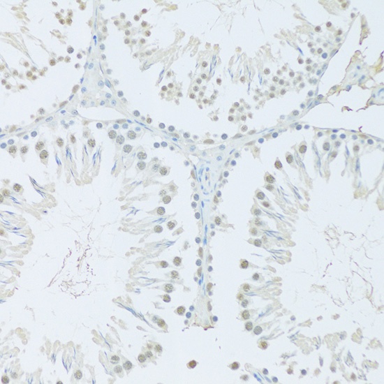
Immunohistochemistry analysis of paraffin-embedded Rat testis using FIP1L1 Rabbit pAb (CAB7138) at dilution of 1:100 (20x lens). Microwave antigen retrieval performed with 0.01M PBS Buffer (pH 7.2) prior to IHC staining.
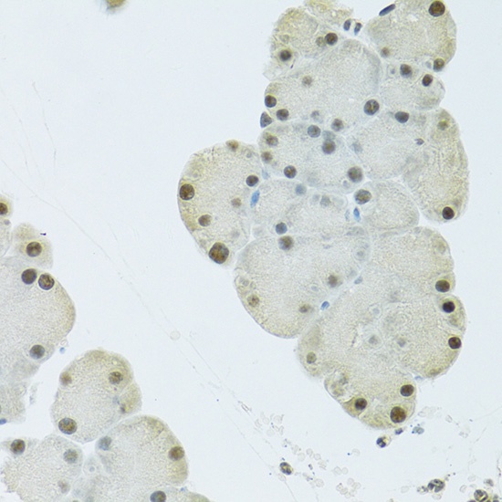
Immunohistochemistry analysis of paraffin-embedded Mouse pancreas using FIP1L1 Rabbit pAb (CAB7138) at dilution of 1:100 (40x lens). Microwave antigen retrieval performed with 0.01M PBS Buffer (pH 7.2) prior to IHC staining.
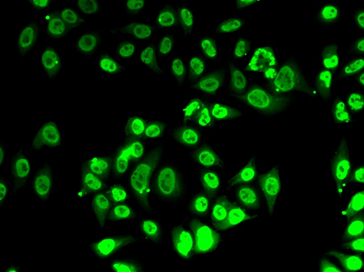
Immunofluorescence analysis of A-549 cells using FIP1L1 Rabbit pAb (CAB7138).