The FLG Antibody (CAB20011) is a high-quality antibody developed for reliable detection and analysis of target proteins. This antibody, raised in rabbits, is highly reactive with human samples and has been validated for use in various applications, including Western blotting and immunohistochemistry.Filaggrin is essential for maintaining the skin barrier function and plays a critical role in protecting against environmental factors and preventing water loss. Mutations in the FLG gene have been associated with skin disorders such as eczema and other inflammatory skin conditions.
This antibody is validated for use in WB, ELISA applications and has demonstrated reactivity against Human, Mouse, Rat samples.
Product Name:
FLG Antibody
SKU:
CAB20011
Size:
20μL, 100μL
Reactivity:
Human, Mouse, Rat
Conjugate:
Unconjugated
Immunogen:
Recombinant protein (or fragment).This information is considered to be commercially sensitive.
The protein encoded by this gene is an intermediate filament-associated protein that aggregates keratin intermediate filaments in mammalian epidermis. It is initially synthesized as a polyprotein precursor, profilaggrin (consisting of multiple filaggrin units of 324 aa each), which is localized in keratohyalin granules, and is subsequently proteolytically processed into individual functional filaggrin molecules. Mutations in this gene are associated with ichthyosis vulgaris.
Purification Method
Affinity purification
Gene ID
2312
RRID
AB_2862917
Buffer Information
Store at -20℃. Avoid freeze / thaw cycles. Buffer: PBS containing 50% glycerol, preserved with proclin300 or sodium azide, pH 7.3.
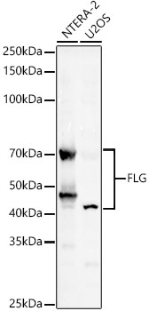
Western blot analysis of various lysates, using FLG Rabbit pAb (CAB20011) at 1:2000 dilution. Secondary antibody: HRP-conjugated Goat anti-Rabbit IgG (H+L) (CABS014) at 1:10000 dilution. Lysates/proteins: 25μg per lane. Blocking buffer: 3% nonfat dry milk in TBST. Detection: ECL Basic Kit (AbGn00020). Exposure time: 180s.
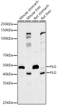
Western blot analysis of various lysates, using FLG Rabbit pAb (CAB20011) at 1:2000 dilution. Secondary antibody: HRP-conjugated Goat anti-Rabbit IgG (H+L) (CABS014) at 1:10000 dilution. Lysates/proteins: 25μg per lane. Blocking buffer: 3% nonfat dry milk in TBST. Detection: ECL Basic Kit (AbGn00020). Exposure time: 180s.