The FLI1 Antibody (CAB5644) is a high-quality antibody developed for reliable detection and analysis of target proteins. This antibody, generated in rabbits, is highly specific to human samples and is validated for use in various applications including Western blot and immunohistochemistry.FLI1, also known as Friend leukemia integration 1 transcription factor, is a key player in the regulation of genes involved in cell growth, differentiation, and apoptosis. Its dysregulation has been linked to various diseases, including leukemia and solid tumors.
This antibody is validated for use in WB, IP, ELISA applications and has demonstrated reactivity against Human, Mouse samples.
Product Name:
FLI1 Antibody
SKU:
CAB5644
Size:
20μL, 100μL
Reactivity:
Human, Mouse
Conjugate:
Unconjugated
Immunogen:
Recombinant protein (or fragment).This information is considered to be commercially sensitive.
0.5μg-4μg antibody for 200μg-400μg extracts of whole cells
ELISA
Recommended starting concentration is 1 μg/mL. Please optimize the concentration based on your specific assay requirements.
Synonyms:
EWSR2, FLI-1, SIC-1, BDPLT21, FLI1
Positive Sample:
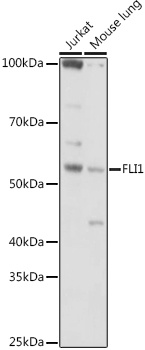
Jurkat, Mouse lung
Cellular Localization:
Nucleus.
Calculated MW:
51kDa
Observed MW:
51kDa
This gene encodes a transcription factor containing an ETS DNA-binding domain. The gene can undergo a t(11;22)(q24;q12) translocation with the Ewing sarcoma gene on chromosome 22, which results in a fusion gene that is present in the majority of Ewing sarcoma cases. An acute lymphoblastic leukemia-associated t(4;11)(q21;q23) translocation involving this gene has also been identified. Alternative splicing results in multiple transcript variants.
Purification Method
Affinity purification
Gene ID
2313
RRID
AB_2766404
Buffer Information
Store at -20℃. Avoid freeze / thaw cycles. Buffer: PBS containing 50% glycerol, preserved with proclin300 or sodium azide, pH 7.3.
Western blot analysis of various lysates using (CAB5644) at 1:1000 dilution. Secondary antibody: HRP-conjugated Goat anti-Rabbit IgG (H+L) (CABS014) at 1:10000 dilution. Lysates/proteins: 25μg per lane. Blocking buffer: 3% nonfat dry milk in TBST. Detection: ECL Basic Kit (AbGn00020). Exposure time: 30s.
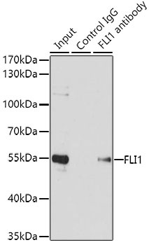
Immunoprecipitation analysis of 200 μg extracts of Jurkat cells using 1 μg FLI1 antibody (CAB5644). Western blot was performed from the immunoprecipitate using FLI1 antibody (CAB5644) at a dilution of 1:1000.