The FLT3 Antibody (CAB12437) is a high-quality antibody developed for reliable detection and analysis of target proteins. This antibody, produced in rabbits, is highly specific for human samples and has been validated for use in Western blot applications.FLT3 is known to play a key role in hematopoiesis and is commonly mutated in acute myeloid leukemia (AML) patients, making it a significant target for cancer research. By targeting the FLT3 protein, researchers can gain insight into the mechanisms of leukemia development and potentially identify new therapeutic strategies for AML treatment.
This antibody is validated for use in WB, IHC-P, ELISA applications and has demonstrated reactivity against Human, Mouse, Rat samples.
Product Name:
FLT3 Antibody
SKU:
CAB12437
Size:
20μL, 100μL
Reactivity:
Human, Mouse, Rat
Conjugate:
Unconjugated
Immunogen:
Synthetic peptide. This information is considered to be commercially sensitive.
Recommended starting concentration is 1 μg/mL. Please optimize the concentration based on your specific assay requirements.
Synonyms:
FLK2, STK1, CD135, FLK-2, FLT3
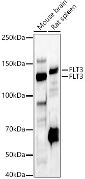
Positive Sample:
Mouse brain, Rat spleen
Cellular Localization:
Endoplasmic Reticulum Lumen, Membrane, Single-Pass Type I Membrane Protein.
Calculated MW:
113kDa
Observed MW:
130kDa/160kDa
This gene encodes a class III receptor tyrosine kinase that regulates hematopoiesis. This receptor is activated by binding of the fms-related tyrosine kinase 3 ligand to the extracellular domain, which induces homodimer formation in the plasma membrane leading to autophosphorylation of the receptor. The activated receptor kinase subsequently phosphorylates and activates multiple cytoplasmic effector molecules in pathways involved in apoptosis, proliferation, and differentiation of hematopoietic cells in bone marrow. Mutations that result in the constitutive activation of this receptor result in acute myeloid leukemia and acute lymphoblastic leukemia.
Purification Method
Affinity purification
Gene ID
2322
RRID
AB_2759279
Buffer Information
Store at -20℃. Avoid freeze / thaw cycles. Buffer: PBS containing 50% glycerol, preserved with proclin300 or sodium azide, pH 7.3.
Western blot analysis of various lysates, using FLT3 Rabbit pAb (CAB12437) at 1:400 dilution. Secondary antibody: HRP-conjugated Goat anti-Rabbit IgG (H+L) (CABS014) at 1:10000 dilution. Lysates/proteins: 25μg per lane. Blocking buffer: 3% nonfat dry milk in TBST. Detection: ECL Enhanced Kit (AbGn00021). Exposure time: 60s.
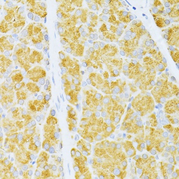
Immunohistochemistry analysis of paraffin-embedded Rat pancreas using FLT3 Rabbit pAb (CAB12437) at dilution of 1:100 (40x lens). Microwave antigen retrieval performed with 0.01M PBS Buffer (pH 7.2) prior to IHC staining.


) ELISA Kit (RBFI00136)")