The FN3KRP Antibody (CAB15512) is a high-quality antibody developed for reliable detection and analysis of target proteins. This antibody, generated in rabbits, is highly specific to human FN3KRP and has been validated for use in Western blot applications. By binding to the FN3KRP protein, this antibody enables accurate detection and analysis in a variety of cell types, making it an essential reagent for investigations into carbohydrate metabolism and related diseases.FN3KRP is a crucial player in the regulation of fructosamine levels, which are implicated in a range of metabolic disorders including diabetes and cardiovascular disease.
This antibody is validated for use in WB, ELISA applications and has demonstrated reactivity against Human, Mouse, Rat samples.
Product Name:
FN3KRP Antibody
SKU:
CAB15512
Size:
20μL, 100μL
Reactivity:
Human, Mouse, Rat
Conjugate:
Unconjugated
Immunogen:
Recombinant protein (or fragment).This information is considered to be commercially sensitive.
Recommended starting concentration is 1 μg/mL. Please optimize the concentration based on your specific assay requirements.
Synonyms:
FN3KL, FN3KRP
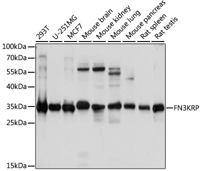
Positive Sample:
293T, U-251MG, MCF7, Mouse brain, Mouse kidney, Mouse lung, Mouse pancreas, Rat spleen, Rat testis
Cellular Localization:
Cytosol.
Calculated MW:
34kDa
Observed MW:
35kDa
A high concentration of glucose can result in non-enzymatic oxidation of proteins by reaction of glucose and lysine residues (glycation). Proteins modified in this way are less active or functional. This gene encodes an enzyme which catalyzes the phosphorylation of psicosamines and ribulosamines compared to the neighboring gene which encodes a highly similar enzyme, fructosamine-3-kinase, which has different substrate specificity. The activity of both enzymes may result in deglycation of proteins to restore their function. Alternative splicing results in multiple transcript variants.
Purification Method
Affinity purification
Gene ID
79672
RRID
AB_2762913
Buffer Information
Store at -20℃. Avoid freeze / thaw cycles. Buffer: PBS with 0.01% thimerosal,50% glycerol,pH7.3.
Western blot analysis of various lysates using FN3KRP Rabbit pAb (CAB15512) at 1:1000 dilution. Secondary antibody: HRP-conjugated Goat anti-Rabbit IgG (H+L) (CABS014) at 1:10000 dilution. Lysates/proteins: 25μg per lane. Blocking buffer: 3% nonfat dry milk in TBST. Detection: ECL Basic Kit (AbGn00020). Exposure time: 5s.