The FOLR1 Antibody (CAB15672) is a high-quality antibody developed for reliable detection and analysis of target proteins. This cell surface protein is involved in the transport of folate into cells, playing a crucial role in cell growth and division. The antibody, generated in rabbits, demonstrates high reactivity with human samples and is validated for use in Western blot applications.By binding specifically to the FOLR1 protein, this antibody enables the detection and analysis of FOLR1 expression in a variety of cell types. Its versatility and reliability make it well-suited for studies in cancer research, as FOLR1 is often overexpressed in cancer cells and has been implicated in tumor growth and progression.
This antibody is validated for use in WB, IHC-P, IF/ICC, ELISA applications and has demonstrated reactivity against Human, Mouse, Rat samples.
Product Name:
FOLR1 Antibody
SKU:
CAB15672
Size:
20μL, 100μL
Reactivity:
Human, Mouse, Rat
Conjugate:
Unconjugated
Immunogen:
Synthetic peptide. This information is considered to be commercially sensitive.
The protein encoded by this gene is a member of the folate receptor family. Members of this gene family bind folic acid and its reduced derivatives, and transport 5-methyltetrahydrofolate into cells. This gene product is a secreted protein that either anchors to membranes via a glycosyl-phosphatidylinositol linkage or exists in a soluble form. Mutations in this gene have been associated with neurodegeneration due to cerebral folate transport deficiency. Due to the presence of two promoters, multiple transcription start sites, and alternative splicing, multiple transcript variants encoding the same protein have been found for this gene.
Purification Method
Affinity purification
Gene ID
2348
RRID
AB_2763081
Buffer Information
Store at -20℃. Avoid freeze / thaw cycles. Buffer: PBS with 0.01% thimerosal,50% glycerol,pH7.3.
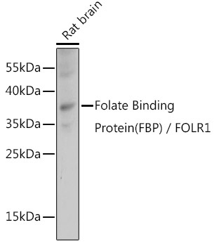
Western blot analysis of lysates from Rat brain, using Folate Binding Protein(FBP) / FOLR1 Rabbit pAb (CAB15672) at 1:1000 dilution. Secondary antibody: HRP-conjugated Goat anti-Rabbit IgG (H+L) (CABS014) at 1:10000 dilution. Lysates/proteins: 25μg per lane. Blocking buffer: 3% nonfat dry milk in TBST. Detection: ECL Basic Kit (AbGn00020). Exposure time: 1s.
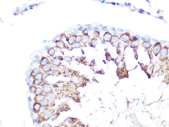
Immunohistochemistry analysis of paraffin-embedded Rat testis using Folate Binding Protein(FBP) / FOLR1 Rabbit pAb (CAB15672) at dilution of 1:100 (40x lens). Microwave antigen retrieval performed with 0.01M Tris/EDTA Buffer (pH 9.0) prior to IHC staining.
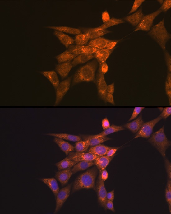
Immunofluorescence analysis of NIH-3T3 cells using Folate Binding Protein(FBP) / FOLR1 Rabbit pAb (CAB15672) at dilution of 1:100 (40x lens). Secondary antibody: Cy3-conjugated Goat anti-Rabbit IgG (H+L) (CABS007) at 1:500 dilution. Blue: DAPI for nuclear staining.
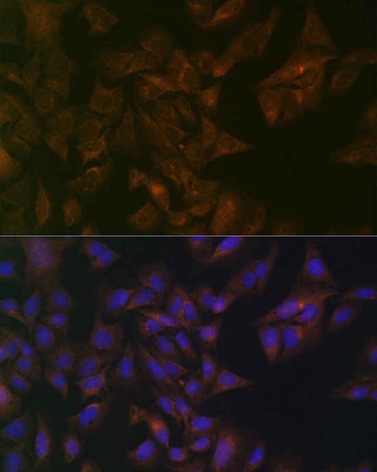
Immunofluorescence analysis of U-2 OS cells using Folate Binding Protein(FBP) / FOLR1 Rabbit pAb (CAB15672) at dilution of 1:100 (40x lens). Secondary antibody: Cy3-conjugated Goat anti-Rabbit IgG (H+L) (CABS007) at 1:500 dilution. Blue: DAPI for nuclear staining.


")
 / FOLR1 Rabbit Monoclonal Antibody (CAB22481)")


