The FREM2 Antibody (CAB15980) is a high-quality antibody developed for reliable detection and analysis of target proteins. This antibody is produced in rabbits and has high reactivity with human samples, making it suitable for use in Western blot applications. By specifically binding to the Frem2 protein, this antibody allows for the detection and analysis of Frem2 in various cell types, making it an essential tool for studies in cell biology and developmental research.Frem2 is a critical protein in the formation of tissues and organs during development, particularly in processes like neural tube closure and heart development.
This antibody is validated for use in WB, IF/ICC, ELISA applications and has demonstrated reactivity against Human, Mouse samples.
Product Name:
FREM2 Antibody
SKU:
CAB15980
Size:
20μL, 100μL
Reactivity:
Human, Mouse
Conjugate:
Unconjugated
Immunogen:
Recombinant protein (or fragment).This information is considered to be commercially sensitive.
Recommended starting concentration is 1 μg/mL. Please optimize the concentration based on your specific assay requirements.
Synonyms:
CRYPTOP, FRASRS2, FREM2
Positive Sample:
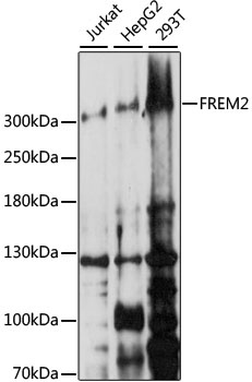
Jurkat, HepG2, 293T
Cellular Localization:
Cell Membrane, Extracellular Side, Single-Pass Type I Membrane Protein.
Calculated MW:
351kDa
Observed MW:
351kDa
This gene encodes an integral membrane protein containing numerous CSPG (chondroitin sulfate proteoglycan element) repeats and Calx-beta domains. The encoded protein localizes to the basement membrane, forming a ternary complex that plays a role in epidermal-dermal interactions. This protein is important for the integrity of skin and renal epithelia. Mutations in this gene are associated with Fraser syndrome.
Purification Method
Affinity purification
Gene ID
341640
RRID
AB_2763419
Buffer Information
Store at -20℃. Avoid freeze / thaw cycles. Buffer: PBS with 0.01% thimerosal,50% glycerol,pH7.3.
Western blot analysis of various lysates using FREM2 Rabbit pAb (CAB15980) at 1:1000 dilution. Secondary antibody: HRP-conjugated Goat anti-Rabbit IgG (H+L) (CABS014) at 1:10000 dilution. Lysates/proteins: 25μg per lane. Blocking buffer: 3% nonfat dry milk in TBST. Detection: ECL Basic Kit (AbGn00020). Exposure time: 150s.
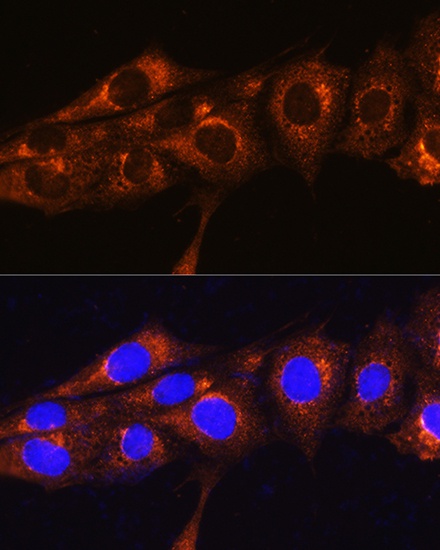
Immunofluorescence analysis of NIH/3T3 cells using FREM2 Rabbit pAb (CAB15980) at dilution of 1:100. Secondary antibody: Cy3-conjugated Goat anti-Rabbit IgG (H+L) (CABS007) at 1:500 dilution. Blue: DAPI for nuclear staining.