The FAH Antibody (CAB6586) is a high-quality antibody developed for reliable detection and analysis of target proteins. This antibody, generated in rabbits, exhibits high specificity and sensitivity for detecting Fumarylacetoacetase in human samples, particularly in Western blot applications. By targeting the Fumarylacetoacetase protein, this antibody enables precise quantification and localization of the enzyme in various cell types.Fumarylacetoacetase plays a critical role in the breakdown of tyrosine and defects in its function have been associated with hereditary tyrosinemia type 1, a rare metabolic disorder.
This antibody is validated for use in WB, IF/ICC, ELISA applications and has demonstrated reactivity against Human, Mouse, Rat samples.
Product Name:
FAH Antibody
SKU:
CAB6586
Size:
20μL, 100μL
Reactivity:
Human, Mouse, Rat
Conjugate:
Unconjugated
Immunogen:
Recombinant protein (or fragment).This information is considered to be commercially sensitive.
Recommended starting concentration is 1 μg/mL. Please optimize the concentration based on your specific assay requirements.
Synonyms:
FAH
Positive Sample:
Mouse liver, Mouse kidney, Rat kidney, Rat liver,
Cellular Localization:
Cytosol, Extracellular Exosome.
Calculated MW:
46kDa
Observed MW:
40-45kDa
Predicted to enable fumarylacetoacetase activity. Predicted to be involved in L-phenylalanine catabolic process; homogentisate catabolic process; and tyrosine catabolic process. Predicted to act upstream of or within arginine catabolic process. Located in extracellular exosome. Implicated in tyrosinemia type I.
Purification Method
Affinity purification
Gene ID
2184
RRID
AB_2767179
Buffer Information
Store at -20℃. Avoid freeze / thaw cycles. Buffer: PBS containing 50% glycerol, preserved with proclin300 or sodium azide, pH 7.3.
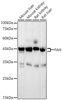
Western blot analysis of various lysates using FAH Rabbit pAb (CAB6586) at 1:10000 dilution. Secondary antibody: HRP-conjugated Goat anti-Rabbit IgG (H+L) (CABS014) at 1:10000 dilution. Lysates / proteins: 25 μg per lane. Blocking buffer: 3 % nonfat dry milk in TBST. Detection: ECL Basic Kit (AbGn00020). Exposure time: 180s.
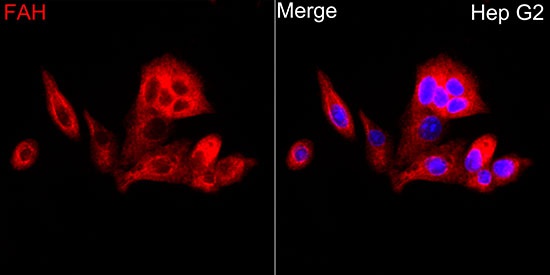
Immunofluorescence analysis of HepG2 cells using FAH Rabbit pAb (CAB6586) at dilution of 1:500 (40x lens). Secondary antibody: Cy3-conjugated Goat anti-Rabbit IgG (H+L) (CABS007) at 1:500 dilution. Blue: DAPI for nuclear staining.