The FXN/Frataxin Antibody (CAB1745) is a high-quality antibody developed for reliable detection and analysis of target proteins. This nuclear gene encodes a mitochondrial protein which belongs to the FRATAXIN family. The protein functions in regulating mitochondrial iron transport and respiration. The expansion of intronic trinucleotide repeat GAA from 8-33 repeats to >90 repeats results in Friedreich ataxia. Alternative splicing results in multiple transcript variants.
This antibody is validated for use in WB, IHC-P, IF/ICC, ELISA applications and has demonstrated reactivity against Human, Mouse, Rat samples.
Product Name:
FXN/Frataxin Antibody
SKU:
CAB1745
Size:
100μL, 20μL
Reactivity:
Human, Mouse, Rat
Conjugate:
Unconjugated
Immunogen:
Recombinant protein (or fragment).This information is considered to be commercially sensitive.
Tested Applications:
WBIHC-PIF/ICCELISA
Recommended Dilution:
WB
1:500 - 1:1000
IHC-P
1:50 - 1:200
IF/ICC
1:50 - 1:200
ELISA
Recommended starting concentration is 1 μg/mL. Please optimize the concentration based on your specific assay requirements.
Synonyms:
FA, X25, CyaY, FARR, FRDA, FXN / Frataxin
Positive Sample:
293T, HepG2, K-562, Mouse heart
Cellular Localization:
Cytoplasm, Mitochondrion.
Calculated MW:
23kDa
Observed MW:
15kDa
This nuclear gene encodes a mitochondrial protein which belongs to the FRATAXIN family. The protein functions in regulating mitochondrial iron transport and respiration. The expansion of intronic trinucleotide repeat GAA from 8-33 repeats to >90 repeats results in Friedreich ataxia. Alternative splicing results in multiple transcript variants.
Purification Method
Affinity purification
Gene ID
2395
RRID
AB_2763791
Buffer Information
Store at -20℃. Avoid freeze / thaw cycles. Buffer: PBS containing 50% glycerol, preserved with proclin300 or sodium azide, pH 7.3.
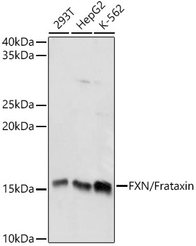
Western blot analysis of various lysates using FXN / Frataxin Rabbit pAb (CAB1745) at 1:1000 dilution. Secondary antibody: HRP-conjugated Goat anti-Rabbit IgG (H+L) (AS014) at 1:10000 dilution. Lysates/proteins: 25μg per lane. Blocking buffer: 3% nonfat dry milk in TBST. Detection: ECL Basic Kit (AbGn00020). Exposure time: 5s.
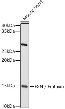
Western blot analysis of lysates from Mouse heart, using FXN / Frataxin Rabbit pAb (CAB1745) at 1:1000 dilution. Secondary antibody: HRP-conjugated Goat anti-Rabbit IgG (H+L) (AS014) at 1:10000 dilution. Lysates/proteins: 25μg per lane. Blocking buffer: 3% nonfat dry milk in TBST. Detection: ECL Basic Kit (AbGn00020). Exposure time: 30s.
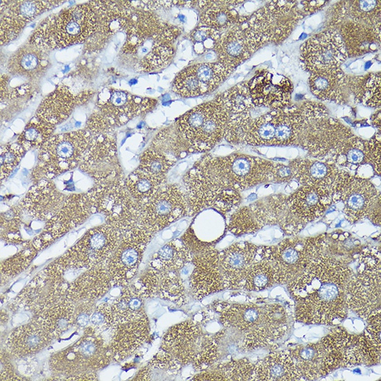
Immunohistochemistry analysis of paraffin-embedded Human liver using FXN / Frataxin Rabbit pAb (CAB1745) at dilution of 1:100 (40x lens). High pressure antigen retrieval performed with 0.01M Citrate buffer (pH 6.0) prior to IHC staining.
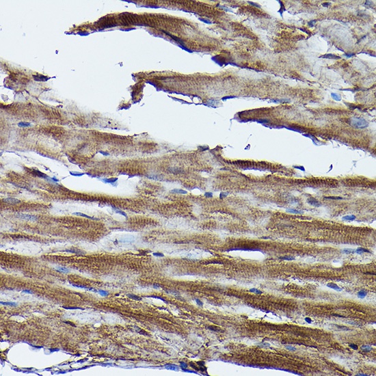
Immunohistochemistry analysis of paraffin-embedded Mouse heart using FXN / Frataxin Rabbit pAb (CAB1745) at dilution of 1:100 (40x lens). High pressure antigen retrieval performed with 0.01M Citrate buffer (pH 6.0) prior to IHC staining.
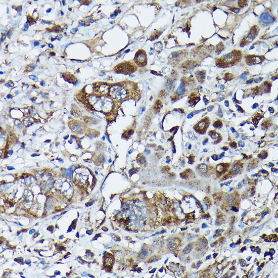
Immunohistochemistry analysis of paraffin-embedded Human liver cancer using FXN / Frataxin Rabbit pAb (CAB1745) at dilution of 1:100 (40x lens). High pressure antigen retrieval performed with 0.01M Citrate buffer (pH 6.0) prior to IHC staining.
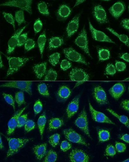
Immunofluorescence analysis of L929 cells using FXN / Frataxin Rabbit pAb (CAB1745) at dilution of 1:100. Secondary antibody: Cy3-conjugated Goat anti-Rabbit IgG (H+L) (AS007) at 1:500 dilution. Blue: DAPI for nuclear staining.
Immunofluorescence analysis of L929 cells using FXN / Frataxin Rabbit pAb (CAB1745) at dilution of 1:100. Secondary antibody: Cy3-conjugated Goat anti-Rabbit IgG (H+L) (AS007) at 1:500 dilution. Blue: DAPI for nuclear staining.
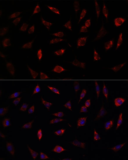
Immunofluorescence analysis of U-2 OS cells using FXN / Frataxin Rabbit pAb (CAB1745) at dilution of 1:100. Secondary antibody: Cy3-conjugated Goat anti-Rabbit IgG (H+L) (AS007) at 1:500 dilution. Blue: DAPI for nuclear staining.
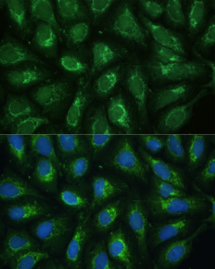
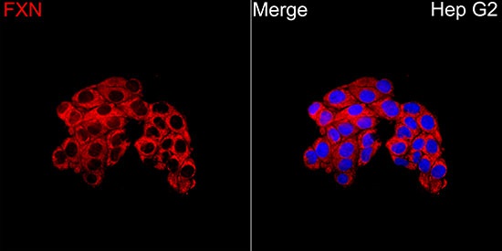
Immunofluorescence analysis of Hep G2 cells using FXN Rabbit pAb (CAB1745) at a dilution of 1:200 (40x lens). Secondary antibody: Cy3-conjugated Goat anti-Rabbit IgG (H+L) (AS007) at 1:500 dilution. Blue: DAPI for nuclear staining.


 ELISA Kit (AEKE02241)")