The G3BP1 Monoclonal Antibody (CAB3968) is a high-quality antibody developed for reliable detection and analysis of target proteins. This antibody, produced using rabbit monoclonal technology, exhibits high reactivity with human samples and is validated for use in immunofluorescence and immunohistochemistry applications. By specifically binding to G3BP, researchers can detect and analyze its expression in various cell types, making it an ideal choice for studies in cellular stress response and RNA regulation.G3BP is known to play a crucial role in cellular stress response pathways, including responses to viral infections, oxidative stress, and heat shock.
This antibody is validated for use in WB, IF/ICC, IP, ELISA applications and has demonstrated reactivity against Human, Mouse, Rat samples.
Product Name:
G3BP1 Monoclonal Antibody
SKU:
CAB3968
Size:
20μL, 100μL
Reactivity:
Human, Mouse, Rat
Clone Number:
ARC0875
Conjugate:
Unconjugated
Immunogen:
Synthetic peptide. This information is considered to be commercially sensitive.
This gene encodes one of the DNA-unwinding enzymes which prefers partially unwound 3'-tailed substrates and can also unwind partial RNA/DNA and RNA/RNA duplexes in an ATP-dependent fashion. This enzyme is a member of the heterogeneous nuclear RNA-binding proteins and is also an element of the Ras signal transduction pathway. It binds specifically to the Ras-GTPase-activating protein by associating with its SH3 domain. Several alternatively spliced transcript variants of this gene have been described, but the full-length nature of some of these variants has not been determined.
Purification Method
Affinity purification
Gene ID
10146
RRID
AB_2863164
Buffer Information
Store at -20℃. Avoid freeze / thaw cycles. Buffer: PBS containing 50% glycerol and 0.05% BSA, preserved with proclin300 or sodium azide, pH 7.3.
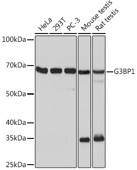
Western blot analysis of various lysates using G3BP1 Rabbit mAb (CAB3968) at 1:1000 dilution. Secondary antibody: HRP-conjugated Goat anti-Rabbit IgG (H+L) (CABS014) at 1:10000 dilution. Lysates/proteins: 25μg per lane. Blocking buffer: 3% nonfat dry milk in TBST. Detection: ECL Basic Kit (AbGn00020). Exposure time: 10s.
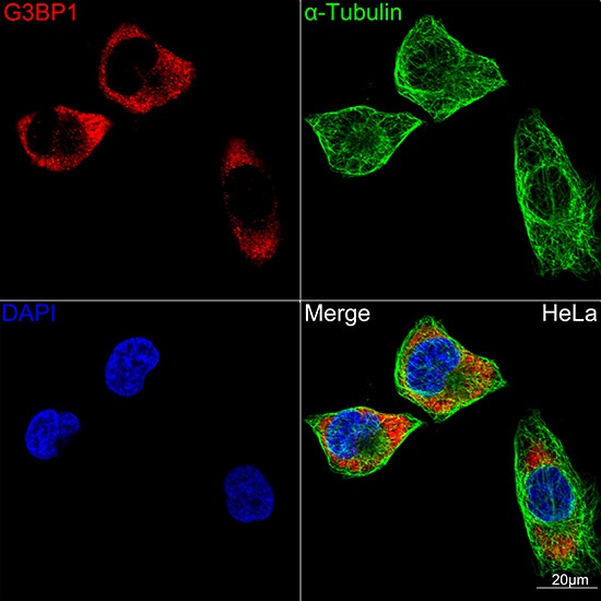
Confocal imaging of HeLa cells using G3BP1 Rabbit mAb (CAB3968,dilution 1:100)(Red). The cells were counterstained with α-Tubulin Mouse mAb (AC012,dilution 1:400) (Green). DAPI was used for nuclear staining (blue). Objective: 100x.
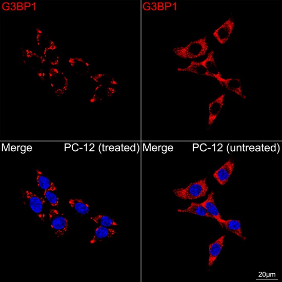
Confocal imaging of PC-12 cells (treated with sodium arsenite) and PC-12 cells (untreated) using G3BP1 Rabbit mAb (CAB3968, dilution 1:100) followed by a further incubation with Cy3 Goat Anti-Rabbit IgG (H+L) (CABS007, dilution 1:500) (Red). DAPI was used for nuclear staining (Blue). Objective: 100x.
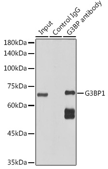
Immunoprecipitation analysis of 300 μg extracts of HeLa cells using 3 μg G3BP1 Rabbit mAb (CAB3968). Western blot was performed from the immunoprecipitate using G3BP1 Rabbit mAb (CAB3968) at a dilution of 1:1000.