The GATA4 Monoclonal Antibody (CAB4306) is a high-quality antibody developed for reliable detection and analysis of target proteins. This antibody, produced in rabbit, exhibits high specificity and sensitivity towards GATA4 in human samples, making it ideal for Western blotting and immunohistochemistry studies.GATA4 plays a crucial role in various biological processes, including cardiac development, endoderm differentiation, and regulation of cell growth. Dysregulation of GATA4 has been implicated in heart defects and certain cancers, highlighting its importance in both development and disease.
This antibody is validated for use in WB, IHC-P, IF/ICC, ELISA applications and has demonstrated reactivity against Human, Mouse, Rat samples.
Product Name:
GATA4 Monoclonal Antibody
SKU:
CAB4306
Size:
20μL, 100μL
Reactivity:
Human, Mouse, Rat
Clone Number:
ARC51718
Conjugate:
Unconjugated
Immunogen:
Synthetic peptide. This information is considered to be commercially sensitive.
Recommended starting concentration is 1 μg/mL. Please optimize the concentration based on your specific assay requirements.
Synonyms:
TOF, ASD2, VSD1, TACHD, GATA4
Positive Sample:
Mouse testis, Rat liver
Cellular Localization:
Nucleus.
Calculated MW:
45kDa
Observed MW:
55kDa
This gene encodes a member of the GATA family of zinc-finger transcription factors. Members of this family recognize the GATA motif which is present in the promoters of many genes. This protein is thought to regulate genes involved in embryogenesis and in myocardial differentiation and function, and is necessary for normal testicular development. Mutations in this gene have been associated with cardiac septal defects. Additionally, alterations in gene expression have been associated with several cancer types. Alternative splicing results in multiple transcript variants.
Purification Method
Affinity purification
Gene ID
2626
RRID
AB_2863232
Buffer Information
Store at -20℃. Avoid freeze / thaw cycles. Buffer: PBS containing 50% glycerol and 0.05% BSA, preserved with proclin300 or sodium azide, pH 7.3.
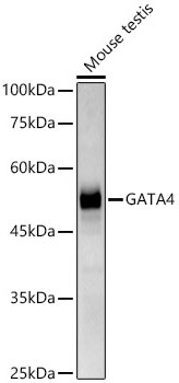
Western blot analysis of lysates from mouse testis, using GATA4 Rabbit mAb (CAB4306) at1:1000 dilution. Secondary antibody: HRP-conjugated Goat anti-Rabbit IgG (H+L) (CABS014) at 1:10000 dilution. Lysates/proteins: 25μg per lane. Blocking buffer: 3% nonfat dry milk in TBST. Detection: ECL Basic Kit (AbGn00020). Exposure time: 180s.
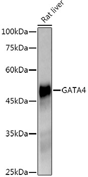
Western blot analysis of lysates from Rat liver, using GATA4 Rabbit mAb (CAB4306) at1:1000 dilution. Secondary antibody: HRP-conjugated Goat anti-Rabbit IgG (H+L) (CABS014) at 1:10000 dilution. Lysates/proteins: 25μg per lane. Blocking buffer: 3% nonfat dry milk in TBST. Detection: ECL Enhanced Kit (AbGn00021). Exposure time: 90s.
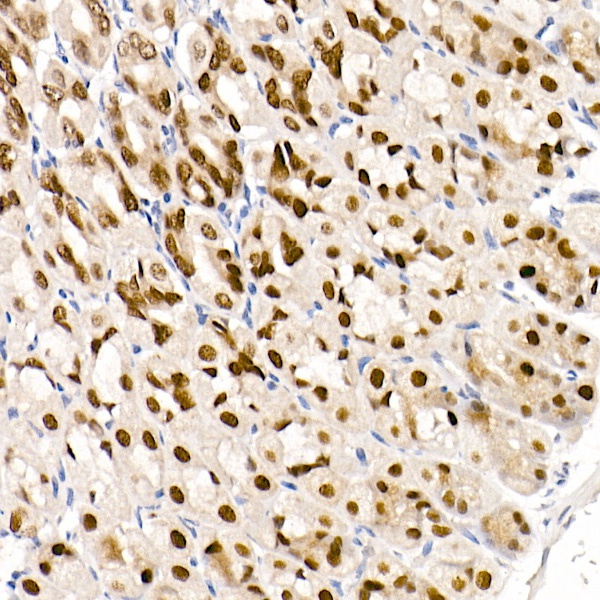
Immunohistochemistry analysis of paraffin-embedded Mouse stomach using GATA4 Rabbit mAb (CAB4306) at dilution of 1:100 (40x lens). High pressure antigen retrieval performed with 0.01M Citrate buffer (pH 6.0) prior to IHC staining.
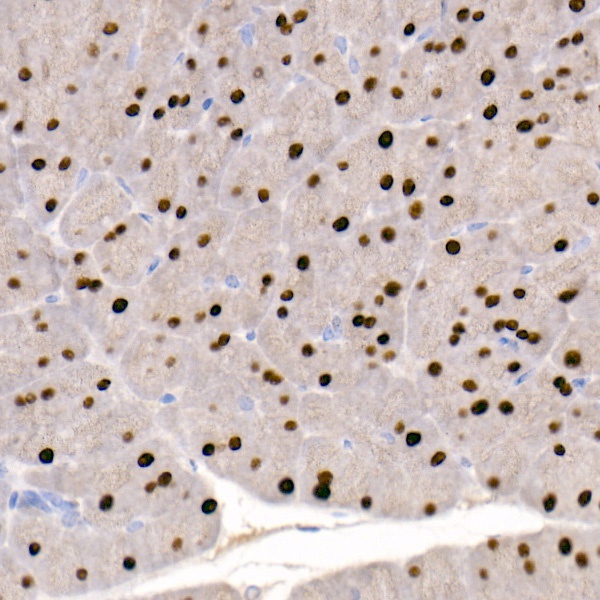
Immunohistochemistry analysis of paraffin-embedded Rat pancreas using GATA4 Rabbit mAb (CAB4306) at dilution of 1:100 (40x lens). High pressure antigen retrieval performed with 0.01M Citrate buffer (pH 6.0) prior to IHC staining.
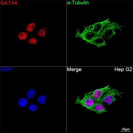
Confocal imaging of Hep G2 cells using GATA4 Rabbit mAb (CAB4306, dilution 1:200) followed by a further incubation with Cy3 Goat Anti-Rabbit IgG (H+L) (CABS007, dilution 1:500) (Red). The cells were counterstained with α-Tubulin Mouse mAb (AC012, dilution 1:400) followed by incubation with ABflo® 488-conjugated Goat Anti-Mouse IgG (H+L) Ab (CABS076, dilution 1:500) (Green). DAPI was used for nuclear staining (Blue). Objective: 100x.