The GCAT Antibody (CAB15802) is a high-quality antibody developed for reliable detection and analysis of target proteins. This antibody, raised in rabbits, shows high specificity for GCAT in human samples and is validated for use in various applications, including Western blotting and immunohistochemistry.GCAT plays a crucial role in the control of purine nucleotide levels, which are essential for DNA and RNA synthesis, energy metabolism, and cell signaling. Dysregulation of GCAT has been implicated in various diseases, including cancer, metabolic disorders, and neurological conditions.
This antibody is validated for use in WB, IHC-P, IF/ICC, ELISA applications and has demonstrated reactivity against Human, Mouse, Rat samples.
Product Name:
GCAT Antibody
SKU:
CAB15802
Size:
20μL, 100μL
Reactivity:
Human, Mouse, Rat
Conjugate:
Unconjugated
Immunogen:
Recombinant protein (or fragment).This information is considered to be commercially sensitive.
Recommended starting concentration is 1 μg/mL. Please optimize the concentration based on your specific assay requirements.
Synonyms:
KBL, GCAT
Positive Sample:
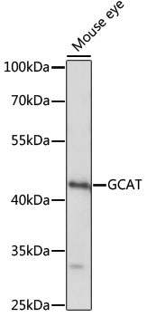
Mouse eye
Cellular Localization:
Mitochondrion, Nucleus.
Calculated MW:
45kDa
Observed MW:
45kDa
The degradation of L-threonine to glycine consists of a two-step biochemical pathway involving the enzymes L-threonine dehydrogenase and 2-amino-3-ketobutyrate coenzyme A ligase. L-Threonine is first converted into 2-amino-3-ketobutyrate by L-threonine dehydrogenase. This gene encodes the second enzyme in this pathway, which then catalyzes the reaction between 2-amino-3-ketobutyrate and coenzyme A to form glycine and acetyl-CoA. The encoded enzyme is considered a class II pyridoxal-phosphate-dependent aminotransferase. Alternate splicing results in multiple transcript variants. A pseudogene of this gene is found on chromosome 14.
Purification Method
Affinity purification
Gene ID
23464
RRID
AB_2763224
Buffer Information
Store at -20℃. Avoid freeze / thaw cycles. Buffer: PBS with 0.01% thimerosal,50% glycerol,pH7.3.
Western blot analysis of lysates from mouse eye, using GCAT Rabbit pAb (CAB15802) at 1:1000 dilution. Secondary antibody: HRP-conjugated Goat anti-Rabbit IgG (H+L) (CABS014) at 1:10000 dilution. Lysates/proteins: 25μg per lane. Blocking buffer: 3% nonfat dry milk in TBST. Detection: ECL Basic Kit (AbGn00020). Exposure time: 10s.
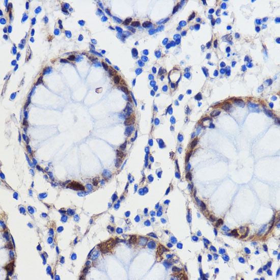
Immunohistochemistry analysis of paraffin-embedded Human colon carcinoma using GCAT Rabbit pAb (CAB15802) at dilution of 1:100 (40x lens). Microwave antigen retrieval performed with 0.01M PBS Buffer (pH 7.2) prior to IHC staining.