The GCSH Antibody (CAB13695) is a high-quality antibody developed for reliable detection and analysis of target proteins. This high-quality antibody, produced in rabbits, exhibits strong reactivity with human samples and is validated for use in Western blotting applications. By targeting the GCSH protein, this antibody allows for precise detection and analysis in a variety of cell types, making it an invaluable resource for studies in metabolism, energy regulation, and mitochondrial function.
This antibody is validated for use in WB, IF/ICC, ELISA applications and has demonstrated reactivity against Human, Mouse, Rat samples.
Product Name:
GCSH Antibody
SKU:
CAB13695
Size:
20μL, 100μL
Reactivity:
Human, Mouse, Rat
Conjugate:
Unconjugated
Immunogen:
Recombinant protein (or fragment).This information is considered to be commercially sensitive.
Recommended starting concentration is 1 μg/mL. Please optimize the concentration based on your specific assay requirements.
Synonyms:
GCE, NKH, GCSH
Positive Sample:
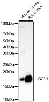
Mouse kidney, Rat kidney
Cellular Localization:
Mitochondrion.
Calculated MW:
19kDa
Observed MW:
15kDa
Degradation of glycine is brought about by the glycine cleavage system, which is composed of four mitochondrial protein components: P protein (a pyridoxal phosphate-dependent glycine decarboxylase), H protein (a lipoic acid-containing protein), T protein (a tetrahydrofolate-requiring enzyme), and L protein (a lipoamide dehydrogenase). The protein encoded by this gene is the H protein, which transfers the methylamine group of glycine from the P protein to the T protein. Defects in this gene are a cause of nonketotic hyperglycinemia (NKH). Two transcript variants, one protein-coding and the other probably not protein-coding,have been found for this gene. Also, several transcribed and non-transcribed pseudogenes of this gene exist throughout the genome.
Purification Method
Affinity purification
Gene ID
2653
RRID
AB_2760555
Buffer Information
Store at -20℃. Avoid freeze / thaw cycles. Buffer: PBS containing 50% glycerol, preserved with proclin300 or sodium azide, pH 7.3.
Western blot analysis of various lysates, using GCSH Rabbit pAb (CAB13695) at 1:700 dilution. Secondary antibody: HRP-conjugated Goat anti-Rabbit IgG (H+L) (CABS014) at 1:10000 dilution. Lysates/proteins: 25μg per lane. Blocking buffer: 3% nonfat dry milk in TBST. Detection: ECL Basic Kit (AbGn00020). Exposure time: 180s.
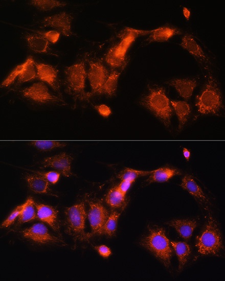
Immunofluorescence analysis of C6 cells using GCSH Rabbit pAb (CAB13695) at dilution of 1:100 (40x lens). Secondary antibody: Cy3-conjugated Goat anti-Rabbit IgG (H+L) (CABS007) at 1:500 dilution. Blue: DAPI for nuclear staining.
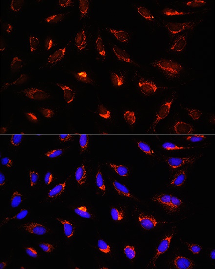
Immunofluorescence analysis of U-2 OS cells using GCSH Rabbit pAb (CAB13695) at dilution of 1:100 (40x lens). Secondary antibody: Cy3-conjugated Goat anti-Rabbit IgG (H+L) (CABS007) at 1:500 dilution. Blue: DAPI for nuclear staining.
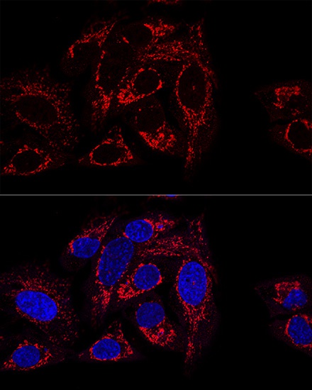
Confocal immunofluorescence analysis of U2OS cells using GCSH Rabbit pAb (CAB13695) at dilution of 1:100 (60x lens). Blue: DAPI for nuclear staining.