The GLUD1 Antibody (CAB7631) is a high-quality antibody developed for reliable detection and analysis of target proteins. This antibody, produced in rabbits, has high specificity and sensitivity for detecting GluD1 in human samples, making it suitable for use in techniques such as Western blotting.The GluD1 protein is involved in excitatory neurotransmission in the brain, making it a key player in processes like learning and memory. Dysregulation of GluD1 has been linked to neurological disorders such as epilepsy and schizophrenia, making it an important target for research in neuroscience and drug development.
This antibody is validated for use in WB, IHC-P, IF/ICC, IP, ELISA applications and has demonstrated reactivity against Human, Mouse, Rat samples.
Product Name:
GLUD1 Antibody
SKU:
CAB7631
Size:
20μL, 100μL
Reactivity:
Human, Mouse, Rat
Conjugate:
Unconjugated
Immunogen:
Recombinant protein (or fragment).This information is considered to be commercially sensitive.
0.5μg-4μg antibody for 200μg-400μg extracts of whole cells
ELISA
Recommended starting concentration is 1 μg/mL. Please optimize the concentration based on your specific assay requirements.
Synonyms:
GDH, GDH1, GLUD, hGDH1, GLUD1
Positive Sample:
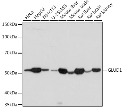
HeLa, HepG2, NIH/3T3, U-251MG, Mouse liver, Mouse brain, Rat liver, Rat brain, Rat kidney
Cellular Localization:
Mitochondrion Matrix.
Calculated MW:
61kDa
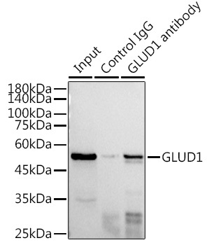
Observed MW:
52kDa
This gene encodes glutamate dehydrogenase, which is a mitochondrial matrix enzyme that catalyzes the oxidative deamination of glutamate to alpha-ketoglutarate and ammonia. This enzyme has an important role in regulating amino acid-induced insulin secretion. It is allosterically activated by ADP and inhibited by GTP and ATP. Activating mutations in this gene are a common cause of congenital hyperinsulinism. Alternative splicing of this gene results in multiple transcript variants. The related glutamate dehydrogenase 2 gene on the human X-chromosome originated from this gene via retrotransposition and encodes a soluble form of glutamate dehydrogenase. Related pseudogenes have been identified on chromosomes 10, 18 and X.
Purification Method
Affinity purification
Gene ID
2746
RRID
AB_2768145
Buffer Information
Store at -20℃. Avoid freeze / thaw cycles. Buffer: PBS with 0.09% Sodium azide,50% glycerol,pH7.3.
Western blot analysis of various lysates using GLUD1 Rabbit pAb (CAB7631) at 1:1000 dilution. Secondary antibody: HRP-conjugated Goat anti-Rabbit IgG (H+L) (CABS014) at 1:10000 dilution. Lysates/proteins: 25μg per lane. Blocking buffer: 3% nonfat dry milk in TBST. Detection: ECL Basic Kit (AbGn00020). Exposure time: 10s.
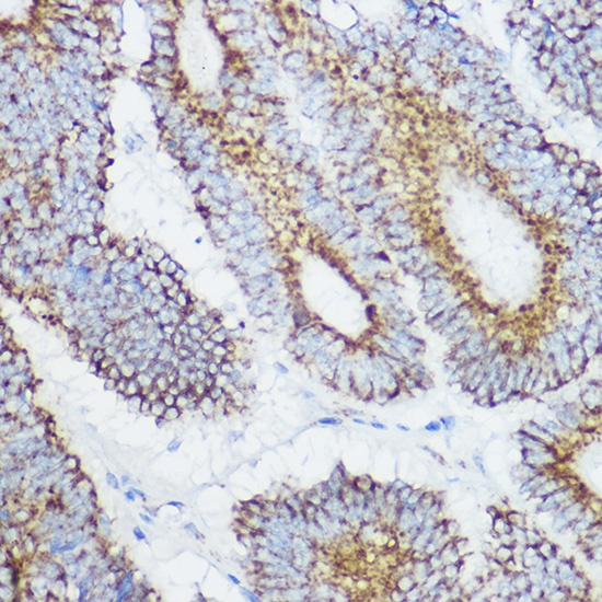
Immunohistochemistry analysis of paraffin-embedded Human colon carcinoma using GLUD1 Rabbit pAb (CAB7631) at dilution of 1:100 (40x lens). High pressure antigen retrieval performed with 0.01M Citrate buffer (pH 6.0) prior to IHC staining.
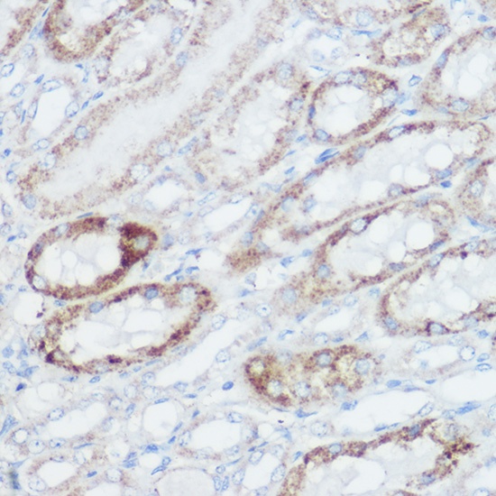
Immunohistochemistry analysis of paraffin-embedded Mouse kidney using GLUD1 Rabbit pAb (CAB7631) at dilution of 1:100 (40x lens). High pressure antigen retrieval performed with 0.01M Citrate buffer (pH 6.0) prior to IHC staining.
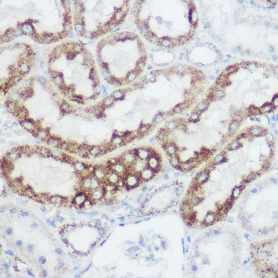
Immunohistochemistry analysis of paraffin-embedded Rat kidney using GLUD1 Rabbit pAb (CAB7631) at dilution of 1:100 (40x lens). High pressure antigen retrieval performed with 0.01M Citrate buffer (pH 6.0) prior to IHC staining.
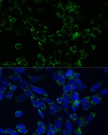
Immunofluorescence analysis of NIH-3T3 cells using GLUD1 Rabbit pAb (CAB7631) at dilution of 1:100. Secondary antibody: Cy3-conjugated Goat anti-Rabbit IgG (H+L) (CABS007) at 1:500 dilution. Blue: DAPI for nuclear staining.
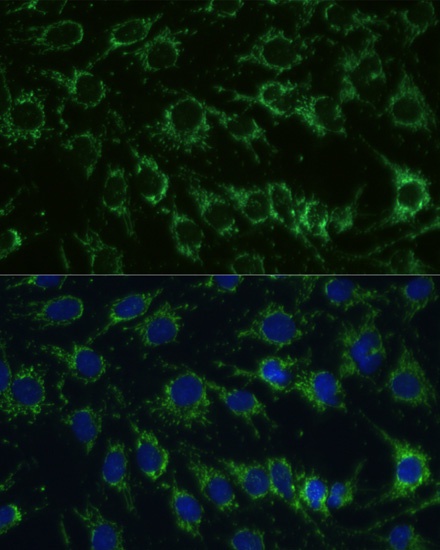
Immunofluorescence analysis of C6 cells using GLUD1 Rabbit pAb (CAB7631) at dilution of 1:100. Secondary antibody: Cy3-conjugated Goat anti-Rabbit IgG (H+L) (CABS007) at 1:500 dilution. Blue: DAPI for nuclear staining.
Western blot analysis of various lysates using GLUD1 Rabbit pAb (CAB7631) at 1:1000 dilution. Secondary antibody: HRP-conjugated Goat anti-Rabbit IgG (H+L) (CABS014) at 1:10000 dilution. Lysates/proteins: 25μg per lane. Blocking buffer: 3% nonfat dry milk in TBST. Detection: ECL Basic Kit (AbGn00020). Exposure time: 10s.