The GLUD1 Monoclonal Antibody (CAB5176) is a high-quality antibody developed for reliable detection and analysis of target proteins. This antibody, raised in rabbits, is highly specific and sensitive to human samples, and is validated for use in Western blot and immunohistochemistry applications.GluD1 is a key player in the regulation of synaptic plasticity and neuronal development, making it a crucial target for studies in neurobiology and neuroscience. The GluD1 Rabbit Monoclonal Antibody binds to the GluD1 protein, enabling precise detection and analysis in various cell types and tissues.
This antibody is validated for use in WB, IHC-P, IF/ICC, ELISA applications and has demonstrated reactivity against Human, Mouse, Rat samples.
Product Name:
GLUD1 Monoclonal Antibody
SKU:
CAB5176
Size:
20μL, 100μL
Reactivity:
Human, Mouse, Rat
Clone Number:
ARC1216
Conjugate:
Unconjugated
Immunogen:
Synthetic peptide. This information is considered to be commercially sensitive.
Recommended starting concentration is 1 μg/mL. Please optimize the concentration based on your specific assay requirements.
Synonyms:
GDH, GDH1, GLUD, hGDH1, GLUD1
Positive Sample:
Jurkat, K-562, U-251MG, Mouse liver, Rat liver
Cellular Localization:
Endoplasmic Reticulum, Mitochondrion.
Calculated MW:
61kDa
Observed MW:
52kDa
This gene encodes glutamate dehydrogenase, which is a mitochondrial matrix enzyme that catalyzes the oxidative deamination of glutamate to alpha-ketoglutarate and ammonia. This enzyme has an important role in regulating amino acid-induced insulin secretion. It is allosterically activated by ADP and inhibited by GTP and ATP. Activating mutations in this gene are a common cause of congenital hyperinsulinism. Alternative splicing of this gene results in multiple transcript variants. The related glutamate dehydrogenase 2 gene on the human X-chromosome originated from this gene via retrotransposition and encodes a soluble form of glutamate dehydrogenase. Related pseudogenes have been identified on chromosomes 10, 18 and X.
Purification Method
Affinity purification
Gene ID
2746
RRID
AB_2863476
Buffer Information
Store at -20℃. Avoid freeze / thaw cycles. Buffer: PBS containing 50% glycerol and 0.05% BSA, preserved with proclin300 or sodium azide, pH 7.3.
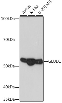
Western blot analysis of various lysates using GLUD1 Rabbit mAb (CAB5176) at 1:1000 dilution. Secondary antibody: HRP-conjugated Goat anti-Rabbit IgG (H+L) (CABS014) at 1:10000 dilution. Lysates/proteins: 25μg per lane. Blocking buffer: 3% nonfat dry milk in TBST. Detection: ECL Basic Kit (AbGn00020). Exposure time: 1s.
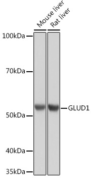
Western blot analysis of various lysates using GLUD1 Rabbit mAb (CAB5176) at 1:1000 dilution. Secondary antibody: HRP-conjugated Goat anti-Rabbit IgG (H+L) (CABS014) at 1:10000 dilution. Lysates/proteins: 25μg per lane. Blocking buffer: 3% nonfat dry milk in TBST. Detection: ECL Basic Kit (AbGn00020). Exposure time: 90s.
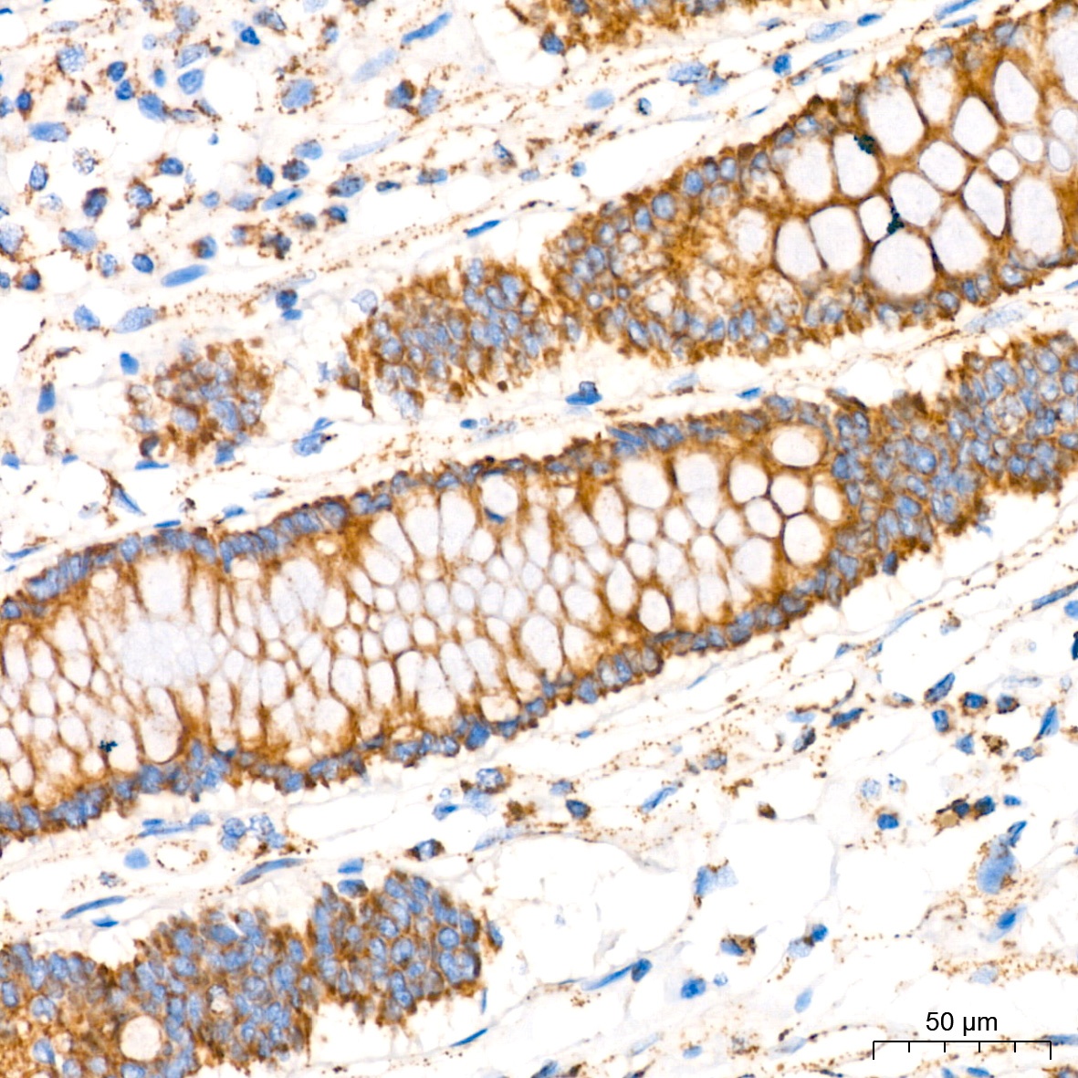
Immunohistochemistry analysis of paraffin-embedded Human colon tissue using GLUD1 Rabbit mAb (CAB5176) at a dilution of 1:200 (40x lens). High pressure antigen retrieval performed with 0.01M Tris-EDTA Buffer (pH 9.0) prior to IHC staining.
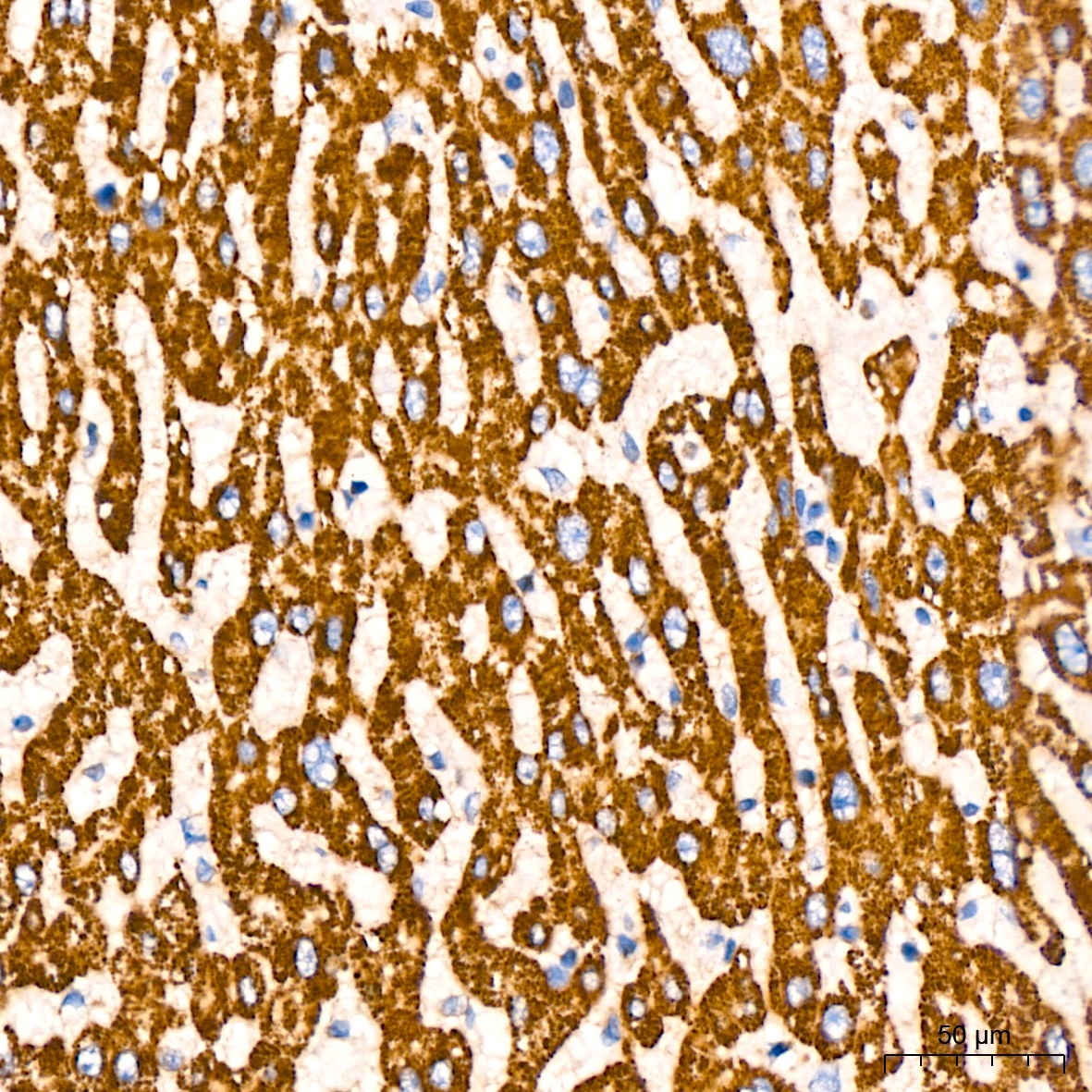
Immunohistochemistry analysis of paraffin-embedded Human liver cancer tissue using GLUD1 Rabbit mAb (CAB5176) at a dilution of 1:200 (40x lens). High pressure antigen retrieval performed with 0.01M Tris-EDTA Buffer (pH 9.0) prior to IHC staining.
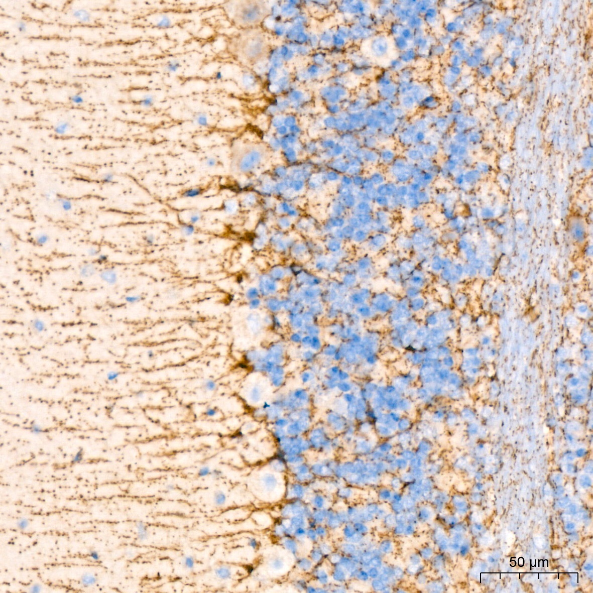
Immunohistochemistry analysis of paraffin-embedded Mouse brain tissue using GLUD1 Rabbit mAb (CAB5176) at a dilution of 1:200 (40x lens). High pressure antigen retrieval performed with 0.01M Tris-EDTA Buffer (pH 9.0) prior to IHC staining.
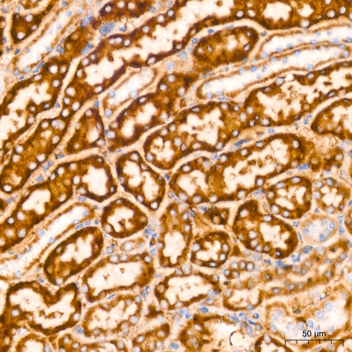
Immunohistochemistry analysis of paraffin-embedded Mouse kidney tissue using GLUD1 Rabbit mAb (CAB5176) at a dilution of 1:200 (40x lens). High pressure antigen retrieval performed with 0.01M Tris-EDTA Buffer (pH 9.0) prior to IHC staining.
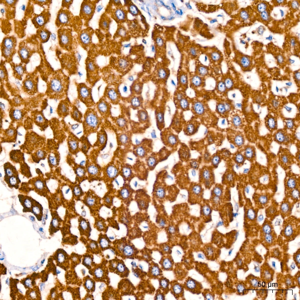
Immunohistochemistry analysis of paraffin-embedded Rat liver tissue using GLUD1 Rabbit mAb (CAB5176) at a dilution of 1:200 (40x lens). High pressure antigen retrieval performed with 0.01M Tris-EDTA Buffer (pH 9.0) prior to IHC staining.
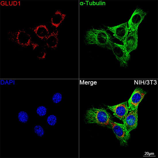
Confocal imaging of NIH/3T3 cells using GLUD1 Rabbit mAb (CAB5176, dilution 1:100) followed by a further incubation with Cy3 Goat Anti-Rabbit IgG (H+L) (CABS007, dilution 1:500) (Red). The cells were counterstained with α-Tubulin Mouse mAb (AC012, dilution 1:400) followed by incubation with ABflo® 488-conjugated Goat Anti-Mouse IgG (H+L) Ab (CABS076, dilution 1:500) (Green). DAPI was used for nuclear staining (Blue). Objective: 100x.