The [KD Validated] GPLD1 Polyclonal Antibody (CAB21781) is a high-quality antibody developed for reliable detection and analysis of target proteins. This antibody, generated in rabbits, demonstrates high specificity and sensitivity in detecting GPLD1 in human samples, making it ideal for use in Western blot and immunohistochemistry applications.GPLD1, also known as phosphatidylglycerophosphatase, is involved in the hydrolysis of glycerophospholipids, contributing to the regulation of lipid levels and immune cell function. Dysregulation of GPLD1 has been implicated in various diseases, including lipid metabolism disorders, immune-related conditions, and certain cancers.
This antibody is validated for use in WB, ELISA applications and has demonstrated reactivity against Human, Mouse samples.
Product Name:
[KD Validated] GPLD1 Polyclonal Antibody
SKU:
CAB21781
Size:
20μL, 100μL
Reactivity:
Human, Mouse
Conjugate:
Unconjugated
Immunogen:
Recombinant protein (or fragment).This information is considered to be commercially sensitive.
Many proteins are tethered to the extracellular face of eukaryotic plasma membranes by a glycosylphosphatidylinositol (GPI) anchor. The GPI-anchor is a glycolipid found on many blood cells. The protein encoded by this gene is a GPI degrading enzyme. Glycosylphosphatidylinositol specific phospholipase D1 hydrolyzes the inositol phosphate linkage in proteins anchored by phosphatidylinositol glycans, thereby releasing the attached protein from the plasma membrane.
Purification Method
Affinity purification
Gene ID
2822
Buffer Information
Store at -20℃. Avoid freeze / thaw cycles. Buffer: PBS containing 50% glycerol, preserved with proclin300 or sodium azide, pH 7.3.
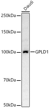
Western blot analysis of lysates from Daudi cells, using [KD Validated] GPLD1 Rabbit pAb (CAB21781) at 1:500 dilution. Secondary antibody: HRP-conjugated Goat anti-Rabbit IgG (H+L) (CABS014) at 1:10000 dilution. Lysates/proteins: 25μg per lane. Blocking buffer: 3% nonfat dry milk in TBST. Detection: ECL Basic Kit (AbGn00020). Exposure time: 1s.
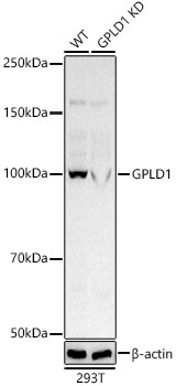
Western blot analysis of lysates from wild type(WT) and GPLD1 knockdown (KD) 293T cells, using [KD Validated] GPLD1 Rabbit pAb (CAB21781) at 1:500 dilution. Secondary antibody: HRP-conjugated Goat anti-Rabbit IgG (H+L) (CABS014) at 1:10000 dilution. Lysates/proteins: 25μg per lane. Blocking buffer: 3% nonfat dry milk in TBST. Detection: ECL Basic Kit (AbGn00020). Exposure time: 1s.
 at 1:500 dilution. Secondary antibody: HRP Goat Anti-Rabbit IgG (H+L) at 1:10000 dilution. Lysates/proteins: 25μg per lane. Blocking buffer: 3% nonfat dry milk in TBST.")
 at 1:500 dilution. Secondary antibody: HRP Goat Anti-Rabbit IgG (H+L) at 1:10000 dilution. Lysates/proteins: 25μg per lane. Blocking buffer: 3% nonfat dry milk in TBST.")

![[KD Validated] COMT Polyclonal Antibody (CAB23600)](https://cdn11.bigcommerce.com/s-h68l9z2lnx/images/stencil/590x590/products/223932/583857/kd-validated-comt-polyclonal-antibody__55967.1701185411.jpg?c=2 "[KD Validated] COMT Polyclonal Antibody (CAB23600)")