The GPLD1 Antibody (CAB6612) is a high-quality antibody developed for reliable detection and analysis of target proteins. Many proteins are tethered to the extracellular face of eukaryotic plasma membranes by a glycosylphosphatidylinositol (GPI) anchor. The GPI-anchor is a glycolipid found on many blood cells. The protein encoded by this gene is a GPI degrading enzyme. Glycosylphosphatidylinositol specific phospholipase D1 hydrolyzes the inositol phosphate linkage in proteins anchored by phosphatidylinositol glycans, thereby releasing the attached protein from the plasma membrane.
This antibody is validated for use in WB, IHC-P, IF/ICC, ELISA applications and has demonstrated reactivity against Human, Mouse, Rat samples.
Product Name:
GPLD1 Antibody
SKU:
CAB6612
Size:
100μL, 20μL
Reactivity:
Human, Mouse, Rat
Conjugate:
Unconjugated
Immunogen:
Recombinant protein (or fragment).This information is considered to be commercially sensitive.
Tested Applications:
WBIHC-PIF/ICCELISA
Recommended Dilution:
WB
1:100 - 1:500
IHC-P
1:50 - 1:200
IF/ICC
1:50 - 1:200
ELISA
Recommended starting concentration is 1 μg/mL. Please optimize the concentration based on your specific assay requirements.
Synonyms:
PLD, GPIPLD, PIGPLD, GPIPLDM, PIGPLD1, GPLD1
Positive Sample:
Mouse liver, Rat liver
Cellular Localization:
Secreted.
Calculated MW:
92kDa
Observed MW:
100kDa
Many proteins are tethered to the extracellular face of eukaryotic plasma membranes by a glycosylphosphatidylinositol (GPI) anchor. The GPI-anchor is a glycolipid found on many blood cells. The protein encoded by this gene is a GPI degrading enzyme. Glycosylphosphatidylinositol specific phospholipase D1 hydrolyzes the inositol phosphate linkage in proteins anchored by phosphatidylinositol glycans, thereby releasing the attached protein from the plasma membrane.
Purification Method
Affinity purification
Gene ID
2822
RRID
AB_2767202
Buffer Information
Store at -20℃. Avoid freeze / thaw cycles. Buffer: PBS with 0.09% Sodium azide,50% glycerol,pH7.3.
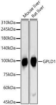
Western blot analysis of various lysates, using GPLD1 Rabbit pAb (CAB6612) at 1:400 dilution. Secondary antibody: HRP-conjugated Goat anti-Rabbit IgG (H+L) (AS014) at 1:10000 dilution. Lysates/proteins: 25μg per lane. Blocking buffer: 3% nonfat dry milk in TBST. Detection: ECL Basic Kit (AbGn00020). Exposure time: 90s.
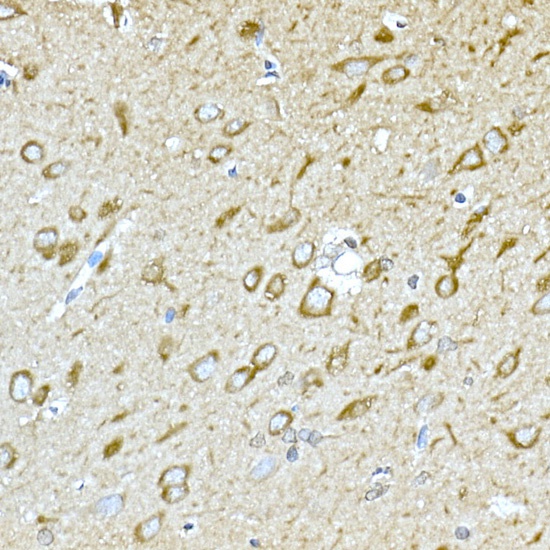
Immunohistochemistry analysis of paraffin-embedded Rat brain using GPLD1 Rabbit pAb (CAB6612) at dilution of 1:100 (40x lens). High pressure antigen retrieval performed with 0.01M Citrate buffer (pH 6.0) prior to IHC staining.
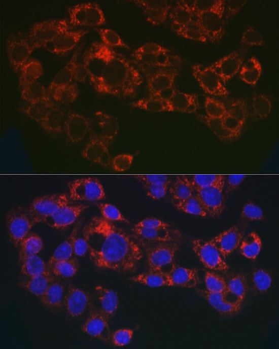
Immunofluorescence analysis of A-549 cells using GPLD1 Rabbit pAb (CAB6612) at dilution of 1:50 (40x lens). Secondary antibody: Cy3-conjugated Goat anti-Rabbit IgG (H+L) (AS007) at 1:500 dilution. Blue: DAPI for nuclear staining.
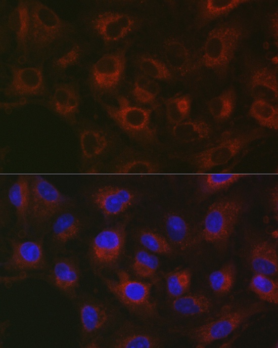
Immunofluorescence analysis of HepG2 cells using GPLD1 Rabbit pAb (CAB6612) at dilution of 1:50 (40x lens). Secondary antibody: Cy3-conjugated Goat anti-Rabbit IgG (H+L) (AS007) at 1:500 dilution. Blue: DAPI for nuclear staining.