The GSTT2B Antibody (CAB15583) is a high-quality antibody developed for reliable detection and analysis of target proteins. This antibody, produced in rabbits, has high specificity for human samples and has been validated for use in Western blot applications. By binding to the GSTT2B protein, this antibody allows for accurate detection and analysis in a variety of cell types, making it ideal for studies in toxicology, pharmacology, and cancer research.
This antibody is validated for use in WB, IF/ICC, ELISA applications and has demonstrated reactivity against Human, Mouse, Rat samples.
Product Name:
GSTT2B Antibody
SKU:
CAB15583
Size:
20μL, 100μL
Reactivity:
Human, Mouse, Rat
Conjugate:
Unconjugated
Immunogen:
Recombinant protein (or fragment).This information is considered to be commercially sensitive.
Recommended starting concentration is 1 μg/mL. Please optimize the concentration based on your specific assay requirements.
Synonyms:
GSTT2P, GSTT2B
Positive Sample:
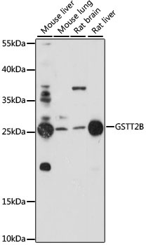
Mouse liver, Mouse lung, Rat brain, Rat liver
Cellular Localization:
Cytoplasm.
Calculated MW:
28kDa
Observed MW:
27kDa
The protein encoded by this gene, glutathione S-transferase (GST) theta 2B (GSTT2B), is a member of a superfamily of proteins that catalyze the conjugation of reduced glutathione to a variety of electrophilic and hydrophobic compounds. Human GSTs can be divided into five main classes: alpha, mu, pi, theta, and zeta. The theta class includes GSTT1, GSTT2, and GSTT2B. GSTT2 and GSTT2B are nearly identical to each other, and share 55% amino acid identity with GSTT1. All three genes may play a role in human carcinogenesis. The GSTT2B gene is a pseudogene in some populations.
Purification Method
Affinity purification
Gene ID
653689
RRID
AB_2762988
Buffer Information
Store at -20℃. Avoid freeze / thaw cycles. Buffer: PBS with 0.01% thimerosal,50% glycerol,pH7.3.
Western blot analysis of various lysates using GSTT2B Rabbit pAb (CAB15583) at 1:1000 dilution. Secondary antibody: HRP-conjugated Goat anti-Rabbit IgG (H+L) (CABS014) at 1:10000 dilution. Lysates/proteins: 25μg per lane. Blocking buffer: 3% nonfat dry milk in TBST. Detection: ECL Basic Kit (AbGn00020). Exposure time: 120s.
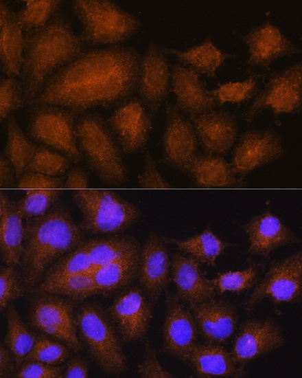
Immunofluorescence analysis of HeLa cells using GSTT2B Rabbit pAb (CAB15583) at dilution of 1:100. Secondary antibody: Cy3-conjugated Goat anti-Rabbit IgG (H+L) (CABS007) at 1:500 dilution. Blue: DAPI for nuclear staining.