The H2-Ab1 Antibody (CAB18658) is a high-quality antibody developed for reliable detection and analysis of target proteins. This rabbit polyclonal antibody is highly specific and reactive with human samples, making it ideal for use in a variety of research applications, particularly in immunology and disease studies.H2-AB1 plays a crucial role in antigen presentation and immune response regulation, making it a key target for investigation in diseases like cancer, autoimmune disorders, and infectious diseases.
This antibody is validated for use in WB, IHC-P, IF/ICC, ELISA applications and has demonstrated reactivity against Human, Mouse, Rat samples.
Product Name:
H2-Ab1 Antibody
SKU:
CAB18658
Size:
20μL, 100μL
Reactivity:
Human, Mouse, Rat
Immunogen:
Recombinant protein (or fragment).This information is considered to be commercially sensitive.
Cell Surface, Early Endosome, External Side Of Plasma Membrane, Golgi Apparatus, Multivesicular Body, Plasma Membrane.
Calculated MW:
30kDa
Observed MW:
30kDa
Enables several functions, including peptide antigen binding activity; protein antigen binding activity; and toxic substance binding activity. Involved in several processes, including B cell affinity maturation; cellular response to interferon-gamma; and positive regulation of T-helper 1 type immune response. Acts upstream of or within antigen processing and presentation of exogenous peptide antigen via MHC class II and immune response. Located in Golgi apparatus; endosome; and external side of plasma membrane. Is integral component of membrane. Part of MHC class II protein complex. Is expressed in lung; metanephros; and thymus primordium. Orthologous to human HLA-DQB2 (major histocompatibility complex, class II, DQ beta 2).
Purification Method
Affinity purification
Gene ID
14961
RRID
AB_2862395
Buffer Information
Store at -20℃. Avoid freeze / thaw cycles. Buffer: PBS containing 50% glycerol, preserved with proclin300 or sodium azide, pH 7.3.
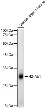
Western blot analysis of lysates from Mouse large intestine, using H2-Ab1 Rabbit pAb (CAB18658) at 1:500 dilution. Secondary antibody: HRP-conjugated Goat anti-Rabbit IgG (H+L) (CABS014) at 1:10000 dilution. Lysates/proteins: 25μg per lane. Blocking buffer: 3% nonfat dry milk in TBST. Detection: ECL Basic Kit (AbGn00020). Exposure time: 20s.
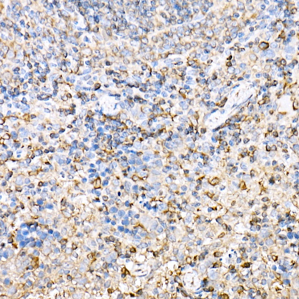
Immunohistochemistry analysis of paraffin-embedded Mouse spleen using H2-Ab1 Rabbit pAb (CAB18658) at dilution of 1:50 (40x lens). High pressure antigen retrieval performed with 0.01M Citrate buffer (pH 6.0) prior to IHC staining.