HA-Tag Monoclonal Antibody is a premium monoclonal that offers outstanding performance and reliability for demanding research applications. Rigorously validated for ELISA, WB, IF, FC, IP, this antibody ensures consistent, reproducible results across multiple experimental platforms. Conveniently packaged in 50ul format to meet your experimental needs. For optimal performance, store at Upon receipt, store at -20°C or -80°C. Avoid repeated freeze. and maintains stability for 12 months. Backed by rigorous quality control testing to ensure superior performance in your critical research applications.
Product Name:
HA-Tag Monoclonal Antibody (MACO0657)
SKU:
MACO0657
Size:
50μl
Isotype:
IgG2b
Host Species:
Mouse
Reactivity:
All
Immunogen:
YPYDVPDYA synthetic peptide conjugate to KLH
Form:
Liquid
Tested Applications:
ELISAWBIFIPFC
Recommended Dilution:
WB 1:5000-1:160000, IF 1:50-1:200, IP 1μg-5μg, FC 1:50-1:200
Western Blot Positive WB detected in: 3 different overexpression lysates with HA tagged All lanes: HA-Tag antibody at 1:1000 Secondary Goat polyclonal to Mouse IgG at 1/10000 dilution Predicted band size: 35, 35, 48 kDa Observed band size: 35, 35, 48 kDa
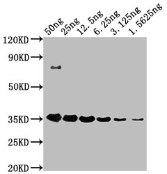
Western Blot Positive WB detected in: HA-tagged fusion protein at 50ng, 25ng, 12.5ng, 6.25ng, 3.125ng, 1.5625ng All lanes: HA-Tag antibody at 1:1000 Secondary Goat polyclonal to Mouse IgG at 1/10000 dilution Predicted band size: 35 kDa Observed band size: 35 kDa
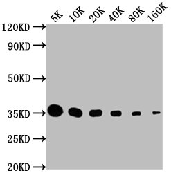
Western Blot Positive WB detected in: HA-tagged fusion protein All lanes: HA-Tag antibody at 1:5000, 1:10000, 1:20000, 1:40000, 1:80000, 1:160000 Secondary Goat polyclonal to Mouse IgG at 1/10000 dilution Predicted band size: 35 kDa Observed band size: 35 kDa
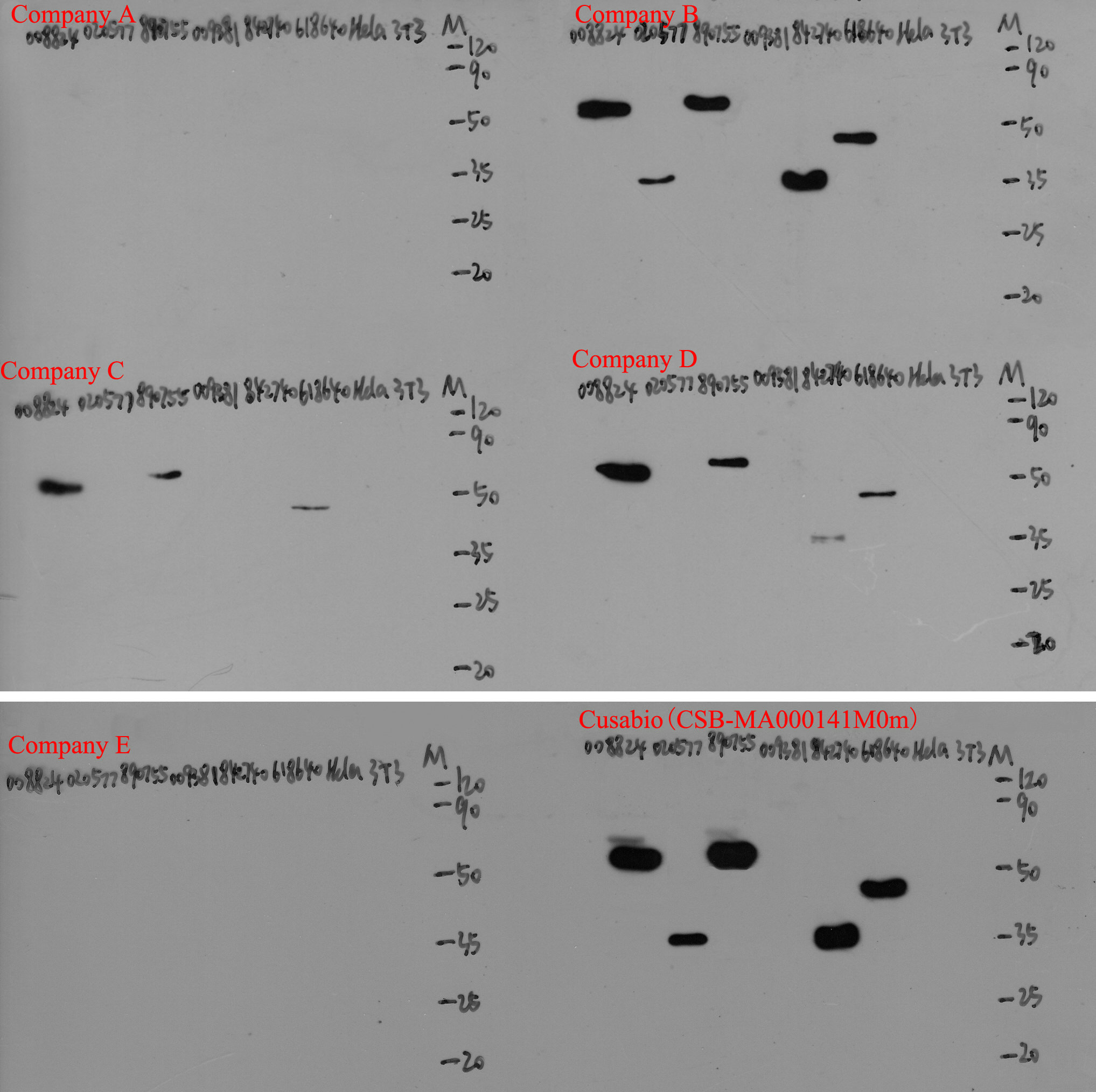
Western Blot Positive WB detected in: 6 different recombinant proteins with HA tagged, Hela whole cell lysate, 3T3 whole cell lysate All lanes: HA-Tag antibody at 1:5000 Secondary Goat polyclonal to Mouse IgG at 1/10000 dilution
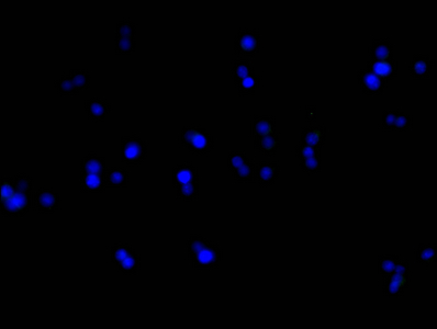
Immunofluorescence staining of 293F cells with MACO0657 at 1:100, counter-stained with DAPI. The cells were blocked in 10% normal Goat Serum and then incubated with the primary antibody overnight at 4°C. The secondary antibody was Alexa Fluor 488-congugated AffiniPure Goat Anti-Mouse IgG(H+L).
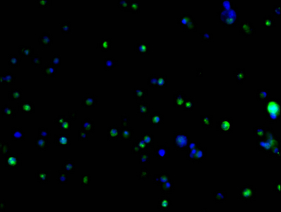
Immunofluorescence staining of 293F transfected cells with MACO0657 at 1:100, counter-stained with DAPI. The cells were blocked in 10% normal Goat Serum and then incubated with the primary antibody overnight at 4°C. The secondary antibody was Alexa Fluor 488-congugated AffiniPure Goat Anti-Mouse IgG(H+L).
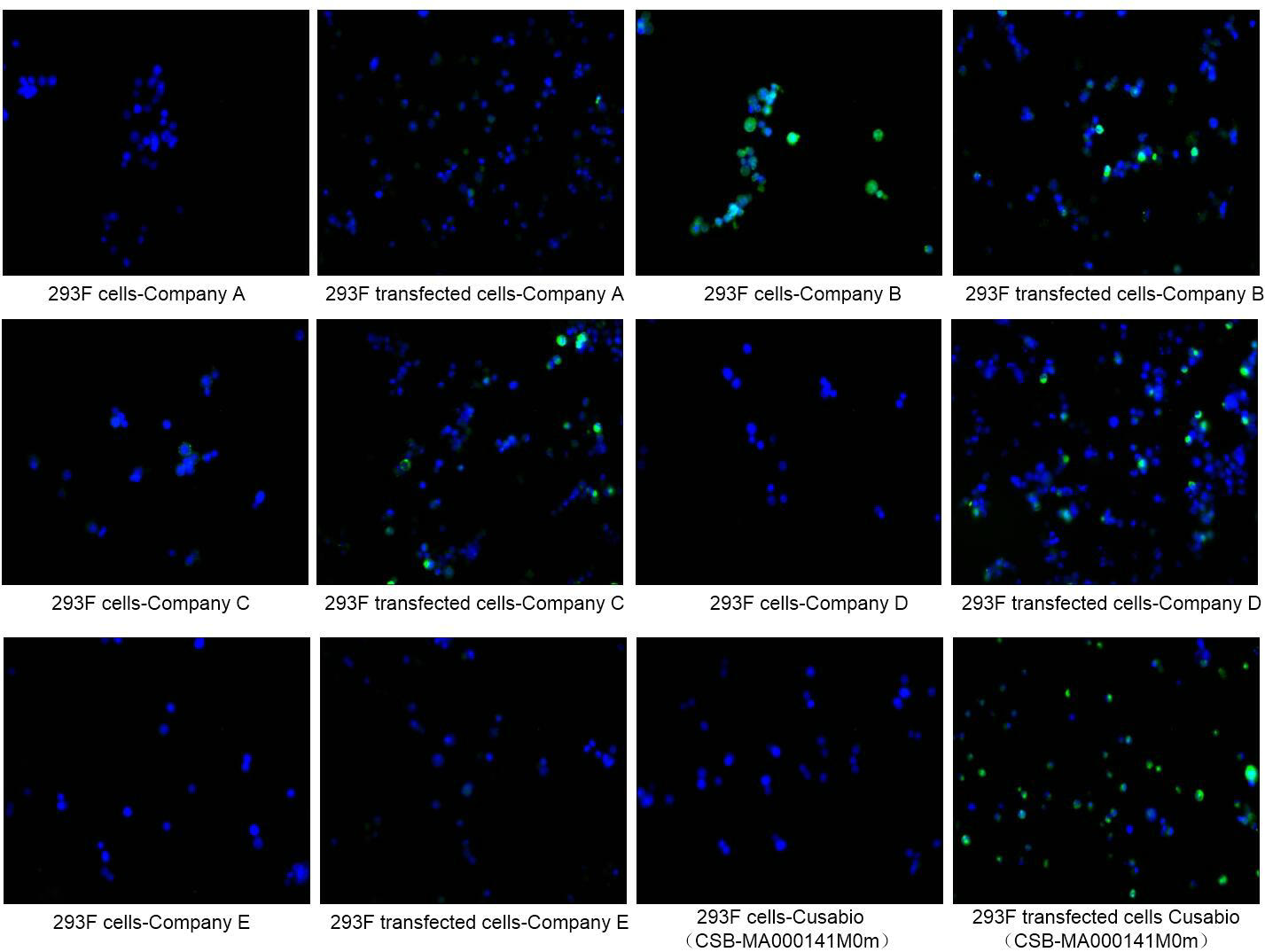
Immunofluorescence staining of 293F cells and 293F transfected cells with Company A, Company B, Company C, Company D, Company E, MACO0657 at 1:100, counter-stained with DAPI. The cells were blocked in 10% normal Goat Serum and then incubated with the primary antibody overnight at 4°C. The secondary antibody was Alexa Fluor 488-congugated AffiniPure Goat Anti-Mouse IgG(H+L).
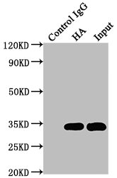
Immunoprecipitating HA-Tag in 293F transfected whole cell lysate Lane 1: Mouse control IgG (1µg) instead of MACO0657 in 293F transfected whole cell lysate. For western blotting, a HRP-conjugated Protein G antibody was used as the secondary antibody (1/2000) Lane 2: MACO0657 (5µg) + 293F transfected whole cell lysate (500µg) Lane 3: 293F transfected whole cell lysate (20µg)
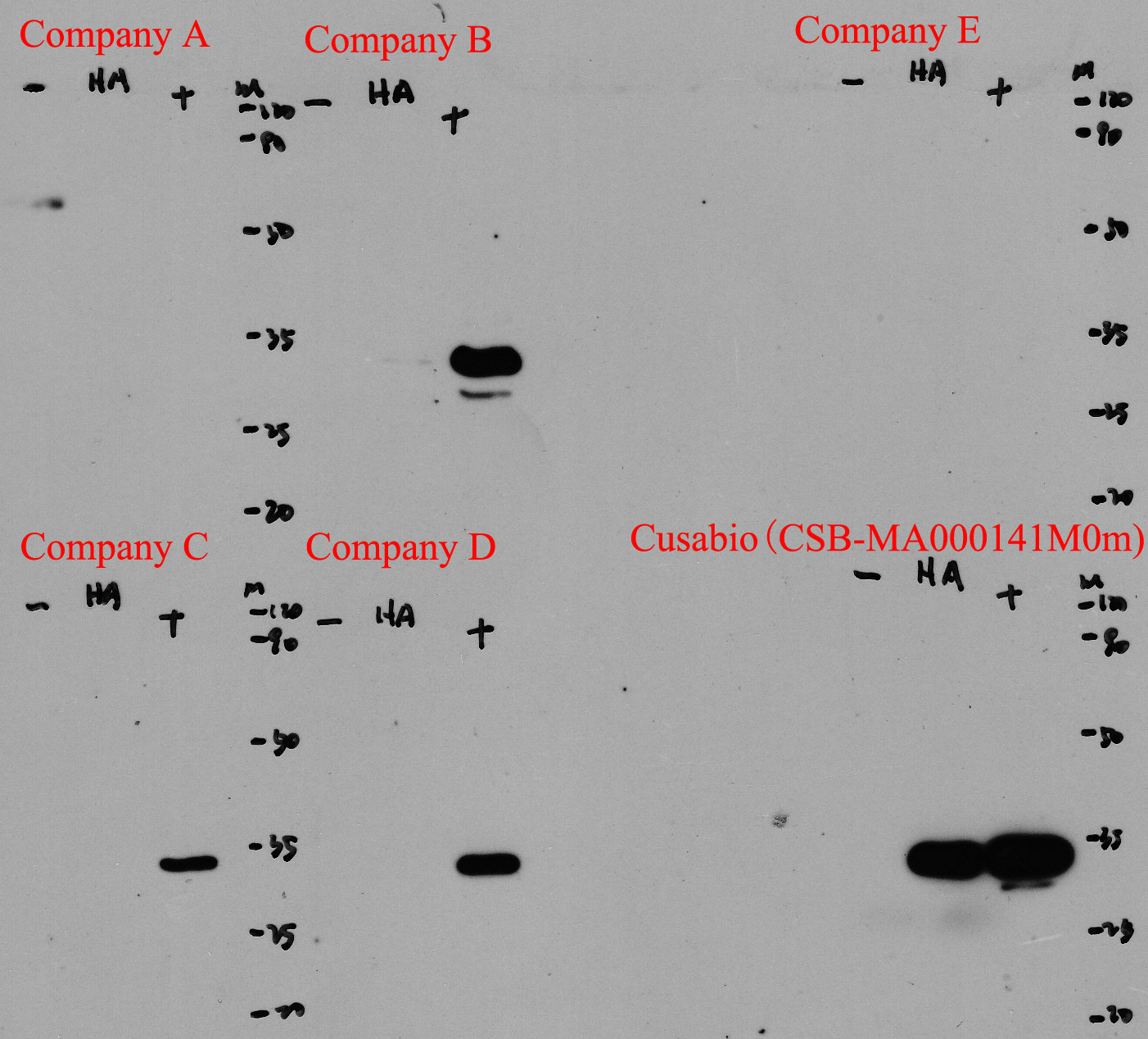
Immunoprecipitating HA-Tag in 293F transfected whole cell lysate Lane 1: Mouse control IgG (1µg) instead of MACO0657 in 293F transfected whole cell lysate. For western blotting, a HRP-conjugated Protein G antibody was used as the secondary antibody (1/2000) Lane 2: Company A (5µg), Company B (5µg),Company C (5µg),Company D (5µg),Company E (5µg),CSB-MA009476A0m (5µg) + 293F transfected whole cell lysate (500µg) Lane 3: 293F transfected whole cell lysate (20µg)
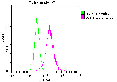
Overlay histogram showing 293F transfected cells stained with MACO0657 (red line) at 1:100. The cells were incubated in 1x PBS /10% normal goat serum to block non-specific protein-protein interactions followed by primary antibody for 1 h at 4°C. The secondary antibody used was FITC goat anti-mouse IgG(H+L) at 1/200 dilution for 1 h at 4°C. Isotype control antibody (green line) was used under the same conditions. Acquisition of >10,000 events was performed.


")
")


