The HAGH Antibody (CAB6615) is a high-quality antibody developed for reliable detection and analysis of target proteins. This antibody, derived from rabbit antibodies, demonstrates high reactivity with human samples and is validated for use in Western blot applications.HAGH, also known as hydroxyacylglutathione hydrolase, is involved in detoxification pathways and has been linked to oxidative stress and aging-related diseases. By targeting the HAGH protein, researchers can investigate its role in cellular metabolism and potentially develop new therapeutic strategies for conditions related to oxidative damage.
This antibody is validated for use in WB, IF/ICC, ELISA applications and has demonstrated reactivity against Human, Mouse, Rat samples.
Product Name:
HAGH Antibody
SKU:
CAB6615
Size:
20μL, 100μL
Reactivity:
Human, Mouse, Rat
Conjugate:
Unconjugated
Immunogen:
Recombinant protein (or fragment).This information is considered to be commercially sensitive.
Recommended starting concentration is 1 μg/mL. Please optimize the concentration based on your specific assay requirements.
Synonyms:
GLO2, GLX2, GLXII, HAGH1, HAGH
Positive Sample:
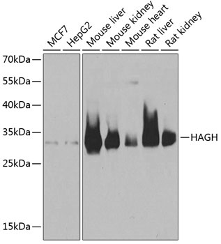
MCF7, HepG2, Mouse liver, Mouse kidney, Mouse heart, Rat liver, Rat kidney
Cellular Localization:
Cytoplasm, Mitochondrion Matrix.
Calculated MW:
34kDa
Observed MW:
34kDa
The enzyme encoded by this gene is classified as a thiolesterase and is responsible for the hydrolysis of S-lactoyl-glutathione to reduced glutathione and D-lactate. Three transcript variants encoding different isoforms have been found for this gene.
Purification Method
Affinity purification
Gene ID
3029
RRID
AB_2767205
Buffer Information
Store at -20℃. Avoid freeze / thaw cycles. Buffer: PBS containing 50% glycerol, preserved with proclin300 or sodium azide, pH 7.3.
Western blot analysis of various lysates using HAGH Rabbit pAb (CAB6615) at 1:1000 dilution. Secondary antibody: HRP-conjugated Goat anti-Rabbit IgG (H+L) (CABS014) at 1:10000 dilution. Lysates/proteins: 25μg per lane. Blocking buffer: 3% nonfat dry milk in TBST. Detection: ECL Basic Kit (AbGn00020). Exposure time: 1s.
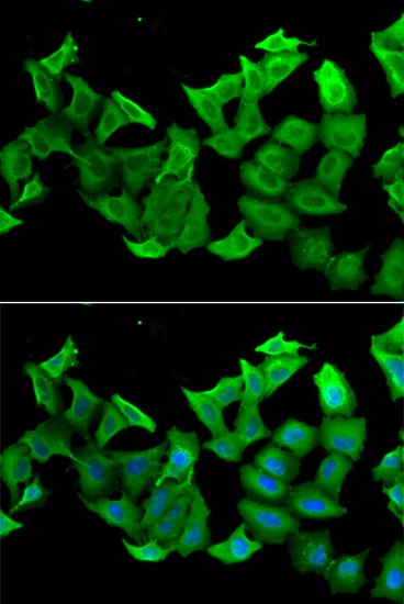
Immunofluorescence analysis of A549 cells using HAGH Rabbit pAb (CAB6615). Secondary antibody: Cy3-conjugated Goat anti-Rabbit IgG (H+L) (CABS007) at 1:500 dilution. Blue: DAPI for nuclear staining.