The HAGH Antibody (CAB6615) is a high-quality antibody developed for reliable detection and analysis of target proteins. The enzyme encoded by this gene is classified as a thiolesterase and is responsible for the hydrolysis of S-lactoyl-glutathione to reduced glutathione and D-lactate. Three transcript variants encoding different isoforms have been found for this gene. RRID AB_2767205 Gene ID 3029 Swiss Prot Synonym GLO2; GLX2; GLXII; HAGH1; HAGH
This antibody is validated for use in WB, IF/ICC, ELISA applications and has demonstrated reactivity against Human, Mouse, Rat samples.
Product Name:
HAGH Antibody
SKU:
CAB6615
Size:
100μL, 20μL
Reactivity:
Human, Mouse, Rat
Clone Number:
-
Conjugate:
Unconjugated
Immunogen:
Recombinant protein (or fragment).This information is considered to be commercially sensitive.
Tested Applications:
WBIF/ICCELISA
Recommended Dilution:
WB
1:500 - 1:2000
IF
/
ICC
1:10 - 1:100
ELISA
Recommended starting concentration is 1 μg/mL. Please optimize the concentration based on your specific assay requirements.
Synonyms:
GLO2, GLX2, GLXII, HAGH1, HAGH
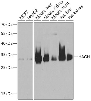
Positive Sample:
MCF7, HepG2, Mouse liver, Mouse kidney, Mouse heart, Rat liver, Rat kidney
Cellular Localization:
Cytoplasm, Mitochondrion Matrix.
Calculated MW:
34kDa
Observed MW:
34kDa
The enzyme encoded by this gene is classified as a thiolesterase and is responsible for the hydrolysis of S-lactoyl-glutathione to reduced glutathione and D-lactate. Three transcript variants encoding different isoforms have been found for this gene. RRID AB_2767205 Gene ID 3029 Swiss Prot Synonym GLO2; GLX2; GLXII; HAGH1; HAGH
Purification Method:
Affinity purification
Gene ID:
3029
RRID:
AB_2767205
Buffer Information:
Store at -20℃. Avoid freeze / thaw cycles. Buffer: PBS containing 50% glycerol, preserved with proclin300 or sodium azide, pH 7.3.
Western blot analysis of various lysates using HAGH Rabbit pAb (CAB6615) at 1:1000 dilution. Secondary antibody: HRP-conjugated Goat anti-Rabbit IgG (H+L) (AS014) at 1:10000 dilution. Lysates/proteins: 25μg per lane. Blocking buffer: 3% nonfat dry milk in TBST. Detection: ECL Basic Kit (AbGn00020). Exposure time: 1s.
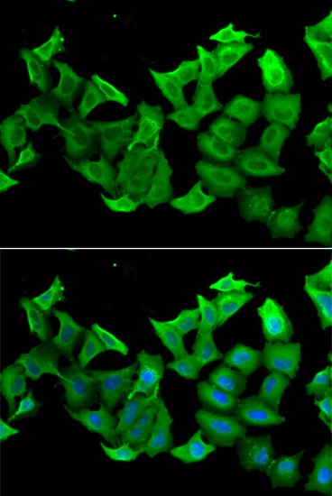
Immunofluorescence analysis of A549 cells using HAGH Rabbit pAb (CAB6615). Secondary antibody: Cy3-conjugated Goat anti-Rabbit IgG (H+L) (AS007) at 1:500 dilution. Blue: DAPI for nuclear staining.